5UP4 | pdb_00005up4

Structure of the HIV-1 Capsid Protein and spacer peptide 1 by Cryo-EM


Experimental Data Snapshot

  • Method: ELECTRON MICROSCOPY
  • Resolution: 9.00 Å
  • Aggregation State: HELICAL ARRAY 
  • Reconstruction Method: HELICAL 

Starting Models: experimental
View more details

wwPDB Validation 3D Report Full Report

Validation slider image for 5UP4

This is version 1.2 of the entry. See complete history

Literature

Quenching protein dynamics interferes with HIV capsid maturation.

Wang, M.Quinn, C.M.Perilla, J.R.Zhang, H.Shirra, R.Hou, G.Byeon, I.J.Suiter, C.L.Ablan, S.Urano, E.Nitz, T.J.Aiken, C.Freed, E.O.Zhang, P.Schulten, K.Gronenborn, A.M.Polenova, T.

(2017) Nat Commun 8: 1779-1779

  • DOI: https://doi.org/10.1038/s41467-017-01856-y
  • Primary Citation Related Structures: 
    5UP4

  • PubMed Abstract: 

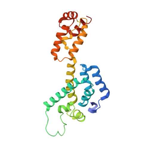
    Maturation of HIV-1 particles encompasses a complex morphological transformation of Gag via an orchestrated series of proteolytic cleavage events. A longstanding question concerns the structure of the C-terminal region of CA and the peptide SP1 (CA-SP1), which represents an intermediate during maturation of the HIV-1 virus. By integrating NMR, cryo-EM, and molecular dynamics simulations, we show that in CA-SP1 tubes assembled in vitro, which represent the features of an intermediate assembly state during maturation, the SP1 peptide exists in a dynamic helix-coil equilibrium, and that the addition of the maturation inhibitors Bevirimat and DFH-055 causes stabilization of a helical form of SP1. Moreover, the maturation-arresting SP1 mutation T8I also induces helical structure in SP1 and further global dynamical and conformational changes in CA. Overall, our results show that dynamics of CA and SP1 are critical for orderly HIV-1 maturation and that small molecules can inhibit maturation by perturbing molecular motions.


  • Organizational Affiliation
    • Department of Chemistry and Biochemistry, University of Delaware, Newark, DE, 19716, USA.

Macromolecule Content 

  • Total Structure Weight: 443.78 kDa 
  • Atom Count: 31,068 
  • Modeled Residue Count: 3,978 
  • Deposited Residue Count: 3,978 
  • Unique protein chains: 1

Macromolecules

Find similar proteins by:|  3D Structure
Entity ID: 1
MoleculeChains  Sequence LengthOrganismDetailsImage
HIV-1 Capsid Protein and spacer peptide 1221Human immunodeficiency virus 1Mutation(s): 0 
Gene Names: gag
UniProt
Find proteins for B6DRA0 (Human immunodeficiency virus type 1)
Explore B6DRA0 
Go to UniProtKB:  B6DRA0
Entity Groups
Sequence Clusters30% Identity50% Identity70% Identity90% Identity95% Identity100% Identity
UniProt GroupB6DRA0
Sequence Annotations
Expand
Reference Sequence

Experimental Data & Validation

Experimental Data

  • Method: ELECTRON MICROSCOPY
  • Resolution: 9.00 Å
  • Aggregation State: HELICAL ARRAY 
  • Reconstruction Method: HELICAL 
EM Software:
TaskSoftware PackageVersion
RECONSTRUCTIONFREALIGN

Structure Validation

View Full Validation Report


Entry History 

& Funding Information

Deposition Data


Funding OrganizationLocationGrant Number
National Institutes of HealthUnited StatesP50 GM082251

Revision History  (Full details and data files)

  • Version 1.0: 2017-12-06
    Type: Initial release
  • Version 1.1: 2017-12-13
    Changes: Refinement description
  • Version 1.2: 2024-11-20
    Changes: Data collection, Database references, Refinement description, Structure summary