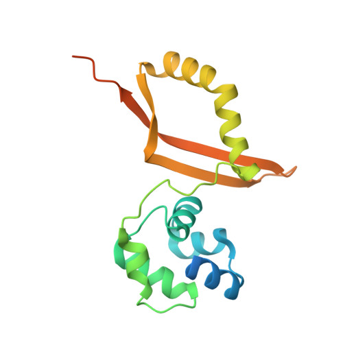
5UK3 | pdb_00005uk3

Crystal structure of cyanase from T. urticae


Experimental Data Snapshot

  • Method: X-RAY DIFFRACTION
  • Resolution: 2.80 Å
  • R-Value Free: 
    0.240 (Depositor), 0.234 (DCC) 
  • R-Value Work: 
    0.221 (Depositor), 0.215 (DCC) 
  • R-Value Observed: 
    0.222 (Depositor) 

Starting Model: experimental
View more details

wwPDB Validation 3D Report Full Report

Validation slider image for 5UK3

This is version 1.3 of the entry. See complete history

Literature

Structural Characterization of a Eukaryotic Cyanase from Tetranychus urticae.

Schlachter, C.R.Klapper, V.Wybouw, N.Radford, T.Van Leeuwen, T.Grbic, M.Chruszcz, M.

(2017) J Agric Food Chem 65: 5453-5462

  • DOI: https://doi.org/10.1021/acs.jafc.7b01333
  • Primary Citation Related Structures: 
    5UK3

  • PubMed Abstract: 

    The two-spotted spider mite Tetranychus urticae is a polyphagous agricultural pest and poses a high risk to global crop production as it is rapidly developing pesticide resistance. Genomic and transcriptomic analysis has revealed the presence of a remarkable cyanase gene in T. urticae and related mite species within the Acariformes lineage. Cyanase catalyzes the detoxification of cyanate and is potentially an attractive protein target for the development of new acaricides. Phylogenetic analysis indicates that within the Acariformes, the cyanase gene originates from a single horizontal gene transfer event, which precedes subsequent speciation. Our structural studies presented here compare and contrast prokaryotic cyanases to T. urticae cyanase, which all form homodecamers and have conserved active site residues, but display different surface areas between homodimers in the overall decameric structure.


  • Organizational Affiliation
    • Department of Chemistry and Biochemistry, University of South Carolina , Columbia, South Carolina 29208, United States.

Macromolecule Content 

  • Total Structure Weight: 202.41 kDa 
  • Atom Count: 10,811 
  • Modeled Residue Count: 1,378 
  • Deposited Residue Count: 1,760 
  • Unique protein chains: 1

Macromolecules

Find similar proteins by:|  3D Structure
Entity ID: 1
MoleculeChains  Sequence LengthOrganismDetailsImage
Uncharacterized protein
A, B, C, D, E
A, B, C, D, E, F, G, H, I, J
176Tetranychus urticaeMutation(s): 0 
UniProt
Find proteins for T1KZQ3 (Tetranychus urticae)
Explore T1KZQ3 
Go to UniProtKB:  T1KZQ3
Entity Groups
Sequence Clusters30% Identity50% Identity70% Identity90% Identity95% Identity100% Identity
UniProt GroupT1KZQ3
Sequence Annotations
Expand
Reference Sequence

Experimental Data & Validation

Experimental Data

  • Method: X-RAY DIFFRACTION
  • Resolution: 2.80 Å
  • R-Value Free:  0.240 (Depositor), 0.234 (DCC) 
  • R-Value Work:  0.221 (Depositor), 0.215 (DCC) 
  • R-Value Observed: 0.222 (Depositor) 
Space Group: P 1 21 1
Unit Cell:
Length ( Å )Angle ( ˚ )
a = 71.074α = 90
b = 81.611β = 100.37
c = 136.466γ = 90
Software Package:
Software NamePurpose
REFMACrefinement
HKL-2000data scaling
MOLREPphasing
HKL-3000phasing

Structure Validation

View Full Validation Report


Entry History 

Deposition Data

Revision History  (Full details and data files)

  • Version 1.0: 2017-06-21
    Type: Initial release
  • Version 1.1: 2017-06-28
    Changes: Database references
  • Version 1.2: 2017-07-26
    Changes: Database references
  • Version 1.3: 2023-10-04
    Changes: Data collection, Database references, Refinement description