Evaluating the Contribution of Transition-State Destabilization to Changes in the Residence Time of Triazole-Based InhA Inhibitors.
Spagnuolo, L.A., Eltschkner, S., Yu, W., Daryaee, F., Davoodi, S., Knudson, S.E., Allen, E.K., Merino, J., Pschibul, A., Moree, B., Thivalapill, N., Truglio, J.J., Salafsky, J., Slayden, R.A., Kisker, C., Tonge, P.J.(2017) J Am Chem Soc 139: 3417-3429
- PubMed: 28151657 Search on PubMedSearch on PubMed Central
- DOI: https://doi.org/10.1021/jacs.6b11148
- Primary Citation Related Structures:
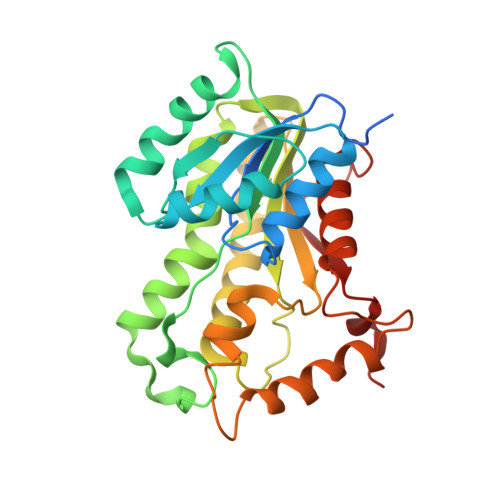
5MTP, 5MTQ, 5MTR, 5UGS, 5UGT, 5UGU - PubMed Abstract:
A critical goal of lead compound selection and optimization is to maximize target engagement while minimizing off-target binding. Since target engagement is a function of both the thermodynamics and kinetics of drug-target interactions, it follows that the structures of both the ground states and transition states on the binding reaction coordinate are needed to rationally modulate the lifetime of the drug-target complex. Previously, we predicted the structure of the rate-limiting transition state that controlled the time-dependent inhibition of the enoyl-ACP reductase InhA. This led to the discovery of a triazole-containing diphenyl ether with an increased residence time on InhA due to transition-state destabilization rather than ground-state stabilization. In the present work, we evaluate the inhibition of InhA by 14 triazole-based diphenyl ethers and use a combination of enzyme kinetics and X-ray crystallography to generate a structure-kinetic relationship for time-dependent binding. We show that the triazole motif slows the rate of formation for the final drug-target complex by up to 3 orders of magnitude. In addition, we identify a novel inhibitor with a residence time on InhA of 220 min, which is 3.5-fold longer than that of the INH-NAD adduct formed by the tuberculosis drug, isoniazid. This study provides a clear example in which the lifetime of the drug-target complex is controlled by interactions in the transition state for inhibitor binding rather than the ground state of the enzyme-inhibitor complex, and demonstrates the important role that on-rates can play in drug-target residence time.
- Institute of Chemical Biology and Drug Discovery, Department of Chemistry, Stony Brook University , Stony Brook, New York 11794-3400, United States.
Organizational Affiliation: