5UGF

Crystal structure of human purine nucleoside phosphorylase (F159Y) mutant complexed with DADMe-ImmG and phosphate


Experimental Data Snapshot

  • Method: X-RAY DIFFRACTION
  • Resolution: 2.20 Å
  • R-Value Free: 0.238 
  • R-Value Work: 0.214 
  • R-Value Observed: 0.215 

wwPDB Validation   3D Report Full Report


Ligand Structure Quality Assessment 


This is version 1.3 of the entry. See complete history


Literature

Catalytic-site design for inverse heavy-enzyme isotope effects in human purine nucleoside phosphorylase.

Harijan, R.K.Zoi, I.Antoniou, D.Schwartz, S.D.Schramm, V.L.

(2017) Proc Natl Acad Sci U S A 114: 6456-6461

  • DOI: https://doi.org/10.1073/pnas.1704786114
  • Primary Citation of Related Structures:  
    5UGF

  • PubMed Abstract: 

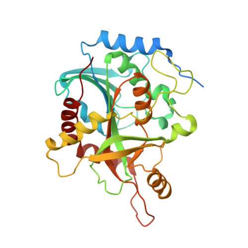
    Heavy-enzyme isotope effects ( 15 N-, 13 C-, and 2 H-labeled protein) explore mass-dependent vibrational modes linked to catalysis. Transition path-sampling (TPS) calculations have predicted femtosecond dynamic coupling at the catalytic site of human purine nucleoside phosphorylase (PNP). Coupling is observed in heavy PNPs, where slowed barrier crossing caused a normal heavy-enzyme isotope effect ( k chem light / k chem heavy > 1.0). We used TPS to design mutant F159Y PNP, predicted to improve barrier crossing for heavy F159Y PNP, an attempt to generate a rare inverse heavy-enzyme isotope effect ( k chem light / k chem heavy < 1.0). Steady-state kinetic comparison of light and heavy native PNPs to light and heavy F159Y PNPs revealed similar kinetic properties. Pre-steady-state chemistry was slowed 32-fold in F159Y PNP. Pre-steady-state chemistry compared heavy and light native and F159Y PNPs and found a normal heavy-enzyme isotope effect of 1.31 for native PNP and an inverse effect of 0.75 for F159Y PNP. Increased isotopic mass in F159Y PNP causes more efficient transition state formation. Independent validation of the inverse isotope effect for heavy F159Y PNP came from commitment to catalysis experiments. Most heavy enzymes demonstrate normal heavy-enzyme isotope effects, and F159Y PNP is a rare example of an inverse effect. Crystal structures and TPS dynamics of native and F159Y PNPs explore the catalytic-site geometry associated with these catalytic changes. Experimental validation of TPS predictions for barrier crossing establishes the connection of rapid protein dynamics and vibrational coupling to enzymatic transition state passage.


  • Organizational Affiliation

    Department of Biochemistry, Albert Einstein College of Medicine, Bronx, NY 10461.


Macromolecules
Find similar proteins by:  (by identity cutoff)  |  3D Structure
Entity ID: 1
MoleculeChains Sequence LengthOrganismDetailsImage
Purine nucleoside phosphorylase
A, B, C, D, E
A, B, C, D, E, F
320Homo sapiensMutation(s): 1 
Gene Names: PNPNP
EC: 2.4.2.1
UniProt & NIH Common Fund Data Resources
Find proteins for P00491 (Homo sapiens)
Explore P00491 
Go to UniProtKB:  P00491
PHAROS:  P00491
GTEx:  ENSG00000198805 
Entity Groups  
Sequence Clusters30% Identity50% Identity70% Identity90% Identity95% Identity100% Identity
UniProt GroupP00491
Sequence Annotations
Expand
  • Reference Sequence
Small Molecules
Ligands 2 Unique
IDChains Name / Formula / InChI Key2D Diagram3D Interactions
IM5
Query on IM5

Download Ideal Coordinates CCD File 
FA [auth D]
LA [auth E]
N [auth A]
OA [auth F]
T [auth B]
FA [auth D],
LA [auth E],
N [auth A],
OA [auth F],
T [auth B],
Y [auth C]
2-amino-7-{[(3R,4R)-3-hydroxy-4-(hydroxymethyl)pyrrolidin-1-yl]methyl}-3,5-dihydro-4H-pyrrolo[3,2-d]pyrimidin-4-one
C12 H17 N5 O3
GSPTUGDLYPMLCQ-SFYZADRCSA-N
PO4
Query on PO4

Download Ideal Coordinates CCD File 
AA [auth D]
BA [auth D]
CA [auth D]
DA [auth D]
EA [auth D]
AA [auth D],
BA [auth D],
CA [auth D],
DA [auth D],
EA [auth D],
G [auth A],
GA [auth E],
H [auth A],
HA [auth E],
I [auth A],
IA [auth E],
J [auth A],
JA [auth E],
K [auth A],
KA [auth E],
L [auth A],
M [auth A],
MA [auth F],
NA [auth F],
O [auth B],
P [auth B],
Q [auth B],
R [auth B],
S [auth B],
U [auth C],
V [auth C],
W [auth C],
X [auth C],
Z [auth D]
PHOSPHATE ION
O4 P
NBIIXXVUZAFLBC-UHFFFAOYSA-K
Binding Affinity Annotations 
IDSourceBinding Affinity
IM5 BindingDB:  5UGF Ki: min: 0.16, max: 7 (nM) from 2 assay(s)
Experimental Data & Validation

Experimental Data

  • Method: X-RAY DIFFRACTION
  • Resolution: 2.20 Å
  • R-Value Free: 0.238 
  • R-Value Work: 0.214 
  • R-Value Observed: 0.215 
  • Space Group: P 21 21 21
Unit Cell:
Length ( Å )Angle ( ˚ )
a = 104.96α = 90
b = 124.27β = 90
c = 136.73γ = 90
Software Package:
Software NamePurpose
REFMACrefinement
PDB_EXTRACTdata extraction
MOSFLMdata reduction
Aimlessdata scaling
PHASERphasing

Structure Validation

View Full Validation Report


Ligand Structure Quality Assessment 


Entry History & Funding Information

Deposition Data


Funding OrganizationLocationGrant Number
National Institutes of Health/National Institute of General Medical Sciences (NIH/NIGMS)United StatesGM068036

Revision History  (Full details and data files)

  • Version 1.0: 2017-06-21
    Type: Initial release
  • Version 1.1: 2017-07-05
    Changes: Database references
  • Version 1.2: 2019-11-27
    Changes: Author supporting evidence
  • Version 1.3: 2023-10-04
    Changes: Data collection, Database references, Refinement description