Perforin proteostasis is regulated through its C2 domain: supra-physiological cell death mediated by T431D-perforin.
Brennan, A.J., Law, R.H.P., Conroy, P.J., Noori, T., Lukoyanova, N., Saibil, H., Yagita, H., Ciccone, A., Verschoor, S., Whisstock, J.C., Trapani, J.A., Voskoboinik, I.(2018) Cell Death Differ 25: 1517-1529
- PubMed: 29416110 Search on PubMedSearch on PubMed Central
- DOI: https://doi.org/10.1038/s41418-018-0057-z
- Primary Citation Related Structures:
5UG6, 5UG7 - PubMed Abstract:
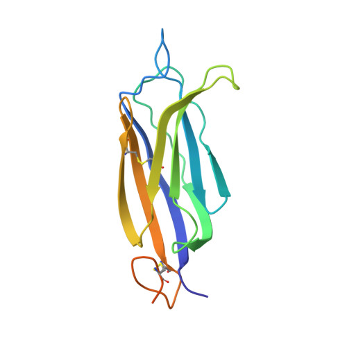
The pore forming, Ca 2+ -dependent protein, perforin, is essential for the function of cytotoxic lymphocytes, which are at the frontline of immune defence against pathogens and cancer. Perforin is a glycoprotein stored in the secretory granules prior to release into the immune synapse. Congenital perforin deficiency causes fatal immune dysregulation, and is associated with various haematological malignancies. At least 50% of pathological missense mutations in perforin result in protein misfolding and retention in the endoplasmic reticulum. However, the regulation of perforin proteostasis remains unexplored. Using a variety of biochemical assays that assess protein stability and acquisition of complex glycosylation, we demonstrated that the binding of Ca 2+ to the C2 domain stabilises perforin and regulates its export from the endoplasmic reticulum to the secretory granules. As perforin is a thermo-labile protein, we hypothesised that by altering its C2 domain it may be possible to improve protein stability. On the basis of the X-ray crystal structure of the perforin C2 domain, we designed a mutation (T431D) in the Ca 2+ binding loop. Mutant perforin displayed markedly enhanced thermal stability and lytic function, despite its trafficking from the endoplasmic reticulum remaining unchanged. Furthermore, by introducing the T431D mutation into A90V perforin, a pathogenic mutation, which results in protein misfolding, we corrected the A90V folding defect and completely restored perforin's cytotoxic function. These results revealed an unexpected role for the Ca 2+ -dependent C2 domain in maintaining perforin proteostasis and demonstrated the possibility of designing perforin with supra-physiological cytotoxic function through stabilisation of the C2 domain.
- Killer Cell Biology Laboratory, Peter MacCallum Cancer Centre, Melbourne, VIC, Australia. amelia.brennan@petermac.org.
Organizational Affiliation: