5UBX | pdb_00005ubx

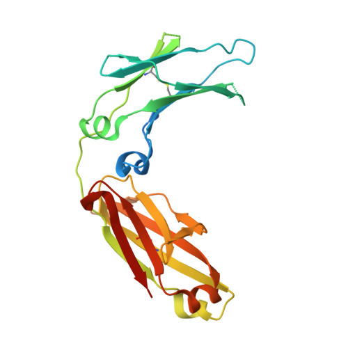
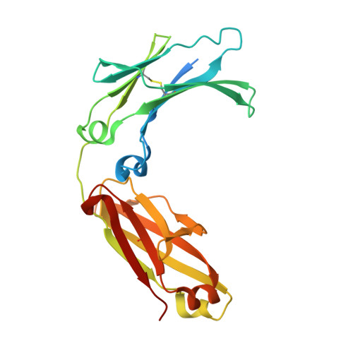
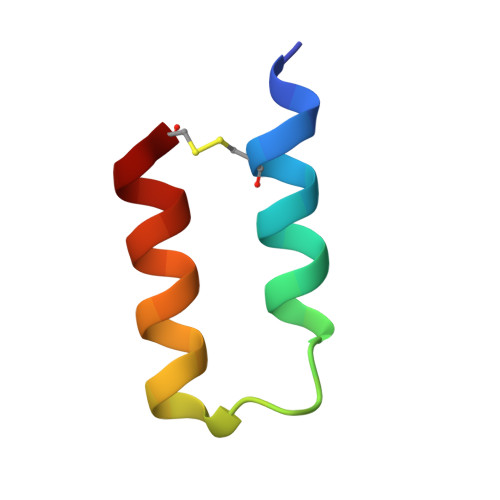
Crystal structure of a mutant mIgG2b Fc heterodimer in complex with Protein A peptide analog Z34C


Experimental Data Snapshot

  • Method: X-RAY DIFFRACTION
  • Resolution: 2.70 Å
  • R-Value Free: 
    0.257 (Depositor), 0.258 (DCC) 
  • R-Value Work: 
    0.209 (Depositor), 0.222 (DCC) 
  • R-Value Observed: 
    0.212 (Depositor) 

Starting Model: experimental
View more details

wwPDB Validation 3D Report Full Report

Validation slider image for 5UBX

This is version 2.2 of the entry. See complete history. 

Literature

Modulation of protein A binding allows single-step purification of mouse bispecific antibodies that retain FcRn binding.

Zwolak, A., Armstrong, A.A., Tam, S.H., Pardinas, J.R., Goulet, D.R., Zheng, S., Brosnan, K., Emmell, E., Luo, J., Gilliland, G.L., Chiu, M.L.

(2017) MAbs 9: 1306-1316

  • DOI: https://doi.org/10.1080/19420862.2017.1375639
  • Primary Citation Related Structures: 
    5UBX

  • PubMed Abstract: 

    The increased number of bispecific antibodies (BsAb) under therapeutic development has resulted in a need for mouse surrogate BsAbs. Here, we describe a one-step method for generating highly pure mouse BsAbs suitable for in vitro and in vivo studies. We identify two mutations in the mouse IgG2a and IgG2b Fc region: one that eliminates protein A binding and one that enhances protein A binding by 8-fold. We show that BsAbs harboring these mutations can be purified from the residual parental monoclonal antibodies in one step using protein A affinity chromatography. The structural basis for the effects of these mutations was analyzed by X-ray crystallography. While the mutation that disrupted protein A binding also inhibited FcRn interaction, a bispecific mutant in which one subunit retained the ability to bind protein A could still interact with FcRn. Pharmacokinetic analysis of the serum half-lives of the mutants showed that the mutant BsAb had a serum half-life comparable to a wild-type Ab. The results describe a rapid method for generating panels of mouse BsAbs that could be used in mouse studies.


  • Organizational Affiliation: 
    • a Biologics Discovery , Janssen Research & Development, LLC , Spring House , PA , USA.

Macromolecule Content 

  • Total Structure Weight: 59.28 kDa 
  • Atom Count: 3,453 
  • Modeled Residue Count: 426 
  • Deposited Residue Count: 500 
  • Unique protein chains: 3

Macromolecules

Find similar proteins by:|  3D Structure
Entity ID: 1
MoleculeChains  Sequence LengthOrganismDetailsImage
Ig gamma-2B chain C region230Mus musculusMutation(s): 4 
Gene Names: Igh-3
UniProt
Find proteins for P01867 (Mus musculus)
Explore P01867 
Go to UniProtKB:  P01867
Entity Groups
Sequence Clusters30% Identity50% Identity70% Identity90% Identity95% Identity100% Identity
UniProt GroupP01867
Sequence Annotations
Expand
Reference Sequence
Find similar proteins by:|  3D Structure
Entity ID: 2
MoleculeChains  Sequence LengthOrganismDetailsImage
Ig gamma-2B chain C region236Mus musculusMutation(s): 2 
Gene Names: Igh-3
UniProt
Find proteins for P01867 (Mus musculus)
Explore P01867 
Go to UniProtKB:  P01867
Entity Groups
Sequence Clusters30% Identity50% Identity70% Identity90% Identity95% Identity100% Identity
UniProt GroupP01867
Glycosylation
Glycosylation Sites: 1Go to GlyGen: P01867-1
Sequence Annotations
Expand
Reference Sequence
Find similar proteins by:|  3D Structure
Entity ID: 3
MoleculeChains  Sequence LengthOrganismDetailsImage
Peptide analog of B-domain from Protein A - Z34CC [auth Z]34Staphylococcus aureusMutation(s): 0 
Entity Groups
Sequence Clusters30% Identity50% Identity70% Identity90% Identity95% Identity100% Identity
Sequence Annotations
Expand
Reference Sequence

Oligosaccharides

Help  
Entity ID: 4
MoleculeChains Length2D Diagram GlycosylationD Interactions
2-acetamido-2-deoxy-beta-D-glucopyranose-(1-2)-alpha-D-mannopyranose-(1-3)-[2-acetamido-2-deoxy-beta-D-glucopyranose-(1-2)-alpha-D-mannopyranose-(1-6)]beta-D-mannopyranose-(1-4)-2-acetamido-2-deoxy-beta-D-glucopyranoseD [auth C]6N/A
Glycosylation Resources
GlyTouCan: G55061AW
GlyCosmos: G55061AW
GlyGen: G55061AW
Entity ID: 5
MoleculeChains Length2D Diagram GlycosylationD Interactions
2-acetamido-2-deoxy-beta-D-glucopyranose-(1-2)-alpha-D-mannopyranose-(1-3)-[2-acetamido-2-deoxy-beta-D-glucopyranose-(1-2)-alpha-D-mannopyranose-(1-6)]beta-D-mannopyranose-(1-4)-2-acetamido-2-deoxy-beta-D-glucopyranose-(1-4)-[alpha-L-fucopyranose-(1-6)]2-acetamido-2-deoxy-beta-D-glucopyranoseE [auth D]8N-Glycosylation
Glycosylation Resources
GlyTouCan: G80858MF
GlyCosmos: G80858MF
GlyGen: G80858MF

Experimental Data & Validation

Experimental Data

  • Method: X-RAY DIFFRACTION
  • Resolution: 2.70 Å
  • R-Value Free:  0.257 (Depositor), 0.258 (DCC) 
  • R-Value Work:  0.209 (Depositor), 0.222 (DCC) 
  • R-Value Observed: 0.212 (Depositor) 
Space Group: P 43 21 2
Unit Cell:
Length ( Å )Angle ( ˚ )
a = 101.49α = 90
b = 101.49β = 90
c = 135.07γ = 90
Software Package:
Software NamePurpose
PHENIXrefinement
XDSdata reduction
XDSdata scaling
PHASERphasing

Structure Validation

View Full Validation Report


Entry History 

Deposition Data

Revision History  (Full details and data files)

  • Version 1.0: 2017-09-20
    Type: Initial release
  • Version 1.1: 2017-10-04
    Changes: Database references
  • Version 1.2: 2017-11-08
    Changes: Database references
  • Version 2.0: 2020-07-29
    Type: Remediation
    Reason: Carbohydrate remediation
    Changes: Advisory, Atomic model, Data collection, Derived calculations, Structure summary
  • Version 2.1: 2023-10-04
    Changes: Data collection, Database references, Derived calculations, Refinement description, Structure summary
  • Version 2.2: 2024-11-20
    Changes: Structure summary