5U7Z

Human acid ceramidase (ASAH1, aCDase) self-activated


Experimental Data Snapshot

  • Method: X-RAY DIFFRACTION
  • Resolution: 2.50 Å
  • R-Value Free: 0.271 
  • R-Value Work: 0.234 
  • R-Value Observed: 0.235 

wwPDB Validation   3D Report Full Report


Ligand Structure Quality Assessment 


This is version 2.0 of the entry. See complete history


Literature

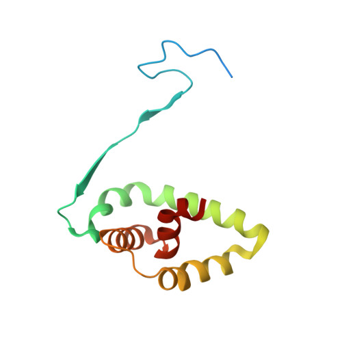
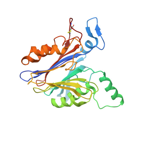
Structural basis for the activation of acid ceramidase.

Gebai, A.Gorelik, A.Li, Z.Illes, K.Nagar, B.

(2018) Nat Commun 9: 1621-1621

  • DOI: https://doi.org/10.1038/s41467-018-03844-2
  • Primary Citation of Related Structures:  
    5U7Z, 5U81, 5U84

  • PubMed Abstract: 

    Acid ceramidase (aCDase, ASAH1) hydrolyzes lysosomal membrane ceramide into sphingosine, the backbone of all sphingolipids, to regulate many cellular processes. Abnormal function of aCDase leads to Farber disease, spinal muscular atrophy with progressive myoclonic epilepsy, and is associated with Alzheimer's, diabetes, and cancer. Here, we present crystal structures of mammalian aCDases in both proenzyme and autocleaved forms. In the proenzyme, the catalytic center is buried and protected from solvent. Autocleavage triggers a conformational change exposing a hydrophobic channel leading to the active site. Substrate modeling suggests distinct catalytic mechanisms for substrate hydrolysis versus autocleavage. A hydrophobic surface surrounding the substrate binding channel appears to be a site of membrane attachment where the enzyme accepts substrates facilitated by the accessory protein, saposin-D. Structural mapping of disease mutations reveals that most would destabilize the protein fold. These results will inform the rational design of aCDase inhibitors and recombinant aCDase for disease therapeutics.


  • Organizational Affiliation

    Department of Biochemistry and Groupe de Recherche Axé sur la Structure des Protéines, McGill University, Montreal, QC, H3G 0B1, Canada.


Macromolecules
Find similar proteins by:  (by identity cutoff)  |  3D Structure
Entity ID: 1
MoleculeChains Sequence LengthOrganismDetailsImage
Acid ceramidase
A, C
129Homo sapiensMutation(s): 0 
UniProt & NIH Common Fund Data Resources
Find proteins for Q13510 (Homo sapiens)
Explore Q13510 
Go to UniProtKB:  Q13510
PHAROS:  Q13510
GTEx:  ENSG00000104763 
Entity Groups  
Sequence Clusters30% Identity50% Identity70% Identity90% Identity95% Identity100% Identity
UniProt GroupQ13510
Sequence Annotations
Expand
  • Reference Sequence
Find similar proteins by:  (by identity cutoff)  |  3D Structure
Entity ID: 2
MoleculeChains Sequence LengthOrganismDetailsImage
Acid ceramidase
B, D
255Homo sapiensMutation(s): 0 
UniProt & NIH Common Fund Data Resources
Find proteins for Q13510 (Homo sapiens)
Explore Q13510 
Go to UniProtKB:  Q13510
PHAROS:  Q13510
GTEx:  ENSG00000104763 
Entity Groups  
Sequence Clusters30% Identity50% Identity70% Identity90% Identity95% Identity100% Identity
UniProt GroupQ13510
Sequence Annotations
Expand
  • Reference Sequence
Oligosaccharides

Help

Entity ID: 3
MoleculeChains Length2D Diagram Glycosylation3D Interactions
beta-D-mannopyranose-(1-4)-2-acetamido-2-deoxy-beta-D-glucopyranose-(1-4)-2-acetamido-2-deoxy-beta-D-glucopyranose
E, F
3N-Glycosylation
Glycosylation Resources
GlyTouCan:  G15407YE
GlyCosmos:  G15407YE
GlyGen:  G15407YE
Small Molecules
Ligands 2 Unique
IDChains Name / Formula / InChI Key2D Diagram3D Interactions
NAG
Query on NAG

Download Ideal Coordinates CCD File 
K [auth B],
L [auth B],
R [auth D],
S [auth D],
T [auth D]
2-acetamido-2-deoxy-beta-D-glucopyranose
C8 H15 N O6
OVRNDRQMDRJTHS-FMDGEEDCSA-N
SO4
Query on SO4

Download Ideal Coordinates CCD File 
G [auth A]
H [auth A]
I [auth A]
J [auth A]
M [auth B]
G [auth A],
H [auth A],
I [auth A],
J [auth A],
M [auth B],
N [auth B],
O [auth B],
P [auth C],
Q [auth C],
U [auth D],
V [auth D],
W [auth D],
X [auth D]
SULFATE ION
O4 S
QAOWNCQODCNURD-UHFFFAOYSA-L
Experimental Data & Validation

Experimental Data

  • Method: X-RAY DIFFRACTION
  • Resolution: 2.50 Å
  • R-Value Free: 0.271 
  • R-Value Work: 0.234 
  • R-Value Observed: 0.235 
  • Space Group: C 1 2 1
Unit Cell:
Length ( Å )Angle ( ˚ )
a = 153.667α = 90
b = 68.312β = 120.72
c = 97.805γ = 90
Software Package:
Software NamePurpose
PHENIXrefinement
HKL-2000data reduction
HKL-2000data scaling
PHASERphasing

Structure Validation

View Full Validation Report


Ligand Structure Quality Assessment 


Entry History 

Deposition Data

Revision History  (Full details and data files)

  • Version 1.0: 2018-03-28
    Type: Initial release
  • Version 1.1: 2018-05-02
    Changes: Data collection, Database references
  • Version 2.0: 2020-07-29
    Type: Remediation
    Reason: Carbohydrate remediation
    Changes: Atomic model, Data collection, Derived calculations, Structure summary