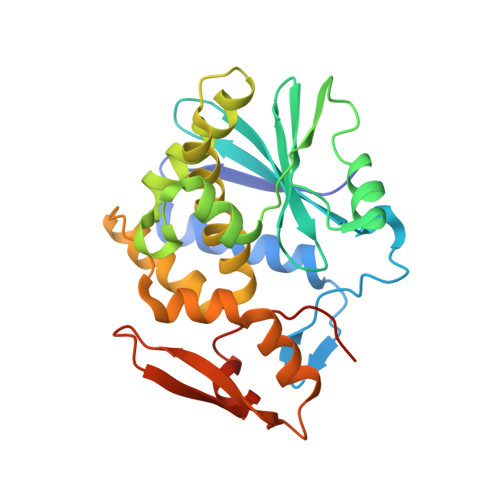
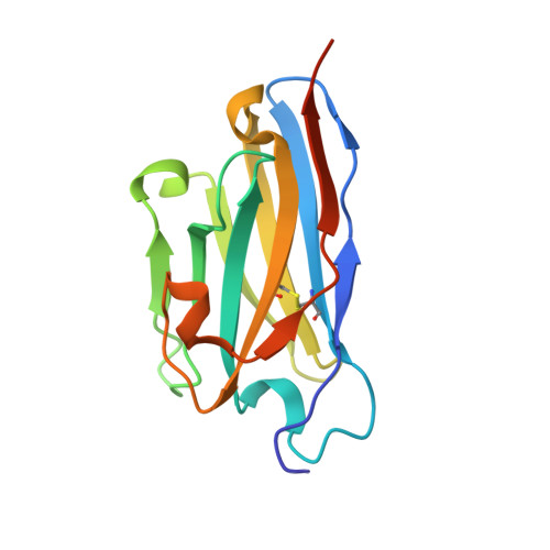
5U4L

RTA-V1C7_G29R-high-salt


Experimental Data Snapshot

  • Method: X-RAY DIFFRACTION
  • Resolution: 2.50 Å
  • R-Value Free: 0.257 
  • R-Value Work: 0.209 
  • R-Value Observed: 0.212 

wwPDB Validation   3D Report Full Report


This is version 1.4 of the entry. See complete history


Literature

Using homology modeling to interrogate binding affinity in neutralization of ricin toxin by a family of single domain antibodies.

Bazzoli, A.Vance, D.J.Rudolph, M.J.Rong, Y.Angalakurthi, S.K.Toth 4th., R.T.Middaugh, C.R.Volkin, D.B.Weis, D.D.Karanicolas, J.Mantis, N.J.

(2017) Proteins 85: 1994-2008

  • DOI: https://doi.org/10.1002/prot.25353
  • Primary Citation of Related Structures:  
    5U4L, 5U4M

  • PubMed Abstract: 

    In this report we investigated, within a group of closely related single domain camelid antibodies (V H Hs), the relationship between binding affinity and neutralizing activity as it pertains to ricin, a fast-acting toxin and biothreat agent. The V1C7-like V H Hs (V1C7, V2B9, V2E8, and V5C1) are similar in amino acid sequence, but differ in their binding affinities and toxin-neutralizing activities. Using the X-ray crystal structure of V1C7 in complex with ricin's enzymatic subunit (RTA) as a template, Rosetta-based homology modeling coupled with energetic decomposition led us to predict that a single pairwise interaction between Arg29 on V5C1 and Glu67 on RTA was responsible for the difference in ricin toxin binding affinity between V1C7, a weak neutralizer, and V5C1, a moderate neutralizer. This prediction was borne out experimentally: substitution of Arg for Gly at position 29 enhanced V1C7's binding affinity for ricin, whereas the reverse (ie, Gly for Arg at position 29) diminished V5C1's binding affinity by >10 fold. As expected, the V5C1 R29G mutant was largely devoid of toxin-neutralizing activity (TNA). However, the TNA of the V1C7 G29R mutant was not correspondingly improved, indicating that in the V1C7 family binding affinity alone does not account for differences in antibody function. V1C7 and V5C1, as well as their respective point mutants, recognized indistinguishable epitopes on RTA, at least at the level of sensitivity afforded by hydrogen-deuterium mass spectrometry. The results of this study have implications for engineering therapeutic antibodies because they demonstrate that even subtle differences in epitope specificity can account for important differences in antibody function.


  • Organizational Affiliation

    Center for Computational Biology, University of Kansas, Lawrence, Kansas, 66045.


Macromolecules
Find similar proteins by:  (by identity cutoff)  |  3D Structure
Entity ID: 1
MoleculeChains Sequence LengthOrganismDetailsImage
Ricin271Ricinus communisMutation(s): 0 
EC: 3.2.2.22
UniProt
Find proteins for P02879 (Ricinus communis)
Explore P02879 
Go to UniProtKB:  P02879
Entity Groups  
Sequence Clusters30% Identity50% Identity70% Identity90% Identity95% Identity100% Identity
UniProt GroupP02879
Sequence Annotations
Expand
  • Reference Sequence
Find similar proteins by:  (by identity cutoff)  |  3D Structure
Entity ID: 2
MoleculeChains Sequence LengthOrganismDetailsImage
V1C7 VHH antibody G29R variant139Vicugna pacosMutation(s): 0 
Entity Groups  
Sequence Clusters30% Identity50% Identity70% Identity90% Identity95% Identity100% Identity
Sequence Annotations
Expand
  • Reference Sequence
Small Molecules
Ligands 3 Unique
IDChains Name / Formula / InChI Key2D Diagram3D Interactions
PO4
Query on PO4

Download Ideal Coordinates CCD File 
C [auth A],
D [auth A],
E [auth A]
PHOSPHATE ION
O4 P
NBIIXXVUZAFLBC-UHFFFAOYSA-K
EDO
Query on EDO

Download Ideal Coordinates CCD File 
K [auth B]1,2-ETHANEDIOL
C2 H6 O2
LYCAIKOWRPUZTN-UHFFFAOYSA-N
CL
Query on CL

Download Ideal Coordinates CCD File 
F [auth A],
G [auth A],
H [auth A],
I [auth A],
J [auth A]
CHLORIDE ION
Cl
VEXZGXHMUGYJMC-UHFFFAOYSA-M
Experimental Data & Validation

Experimental Data

  • Method: X-RAY DIFFRACTION
  • Resolution: 2.50 Å
  • R-Value Free: 0.257 
  • R-Value Work: 0.209 
  • R-Value Observed: 0.212 
  • Space Group: P 32 2 1
Unit Cell:
Length ( Å )Angle ( ˚ )
a = 64.97α = 90
b = 64.97β = 90
c = 215.968γ = 120
Software Package:
Software NamePurpose
SCALEPACKdata scaling
PHENIXrefinement
PDB_EXTRACTdata extraction
HKL-2000data reduction
PHASERphasing

Structure Validation

View Full Validation Report


Entry History & Funding Information

Deposition Data


Funding OrganizationLocationGrant Number
National Institutes of Health/National Institute Of Allergy and Infectious Diseases (NIH/NIAID)United StatesHHSN272201400021C

Revision History  (Full details and data files)

  • Version 1.0: 2017-12-13
    Type: Initial release
  • Version 1.1: 2018-01-17
    Changes: Author supporting evidence
  • Version 1.2: 2018-12-26
    Changes: Data collection, Database references
  • Version 1.3: 2019-12-11
    Changes: Author supporting evidence
  • Version 1.4: 2023-10-04
    Changes: Data collection, Database references, Refinement description