5TXG | pdb_00005txg

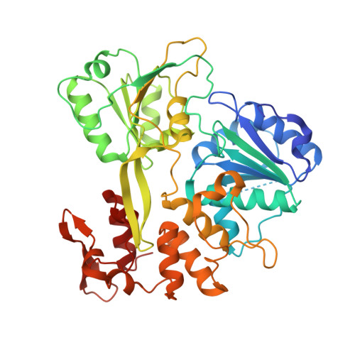
Crystal structure of the Zika virus NS3 helicase.


Experimental Data Snapshot

  • Method: X-RAY DIFFRACTION
  • Resolution: 2.05 Å
  • R-Value Free: 
    0.247 (Depositor), 0.269 (DCC) 
  • R-Value Work: 
    0.184 (Depositor) 
  • R-Value Observed: 
    0.187 (Depositor) 

Starting Model: experimental
View more details

wwPDB Validation 3D Report Full Report

Validation slider image for 5TXG

This is version 1.1 of the entry. See complete history

Macromolecule Content 

  • Total Structure Weight: 50.75 kDa 
  • Atom Count: 3,693 
  • Modeled Residue Count: 432 
  • Deposited Residue Count: 449 
  • Unique protein chains: 1

Macromolecules

Find similar proteins by:|  3D Structure
Entity ID: 1
MoleculeChains  Sequence LengthOrganismDetailsImage
NS3 helicase449Zika virusMutation(s): 0 
UniProt
Find proteins for A0A0X8GJ44 (Zika virus)
Explore A0A0X8GJ44 
Go to UniProtKB:  A0A0X8GJ44
Entity Groups
Sequence Clusters30% Identity50% Identity70% Identity90% Identity95% Identity100% Identity
UniProt GroupA0A0X8GJ44
Sequence Annotations
Expand
Reference Sequence

Experimental Data & Validation

Experimental Data

  • Method: X-RAY DIFFRACTION
  • Resolution: 2.05 Å
  • R-Value Free:  0.247 (Depositor), 0.269 (DCC) 
  • R-Value Work:  0.184 (Depositor) 
  • R-Value Observed: 0.187 (Depositor) 
Space Group: P 1 21 1
Unit Cell:
Length ( Å )Angle ( ˚ )
a = 51.639α = 90
b = 69.855β = 93.41
c = 58.461γ = 90
Software Package:
Software NamePurpose
REFMACrefinement
HKL-3000data reduction
HKL-3000data scaling
PHASERphasing

Structure Validation

View Full Validation Report


Entry History 

Revision History  (Full details and data files)

  • Version 1.0: 2016-12-14
    Type: Initial release
  • Version 1.1: 2023-10-04
    Changes: Data collection, Database references, Refinement description