Computational design of environmental sensors for the potent opioid fentanyl.
Bick, M.J., Greisen, P.J., Morey, K.J., Antunes, M.S., La, D., Sankaran, B., Reymond, L., Johnsson, K., Medford, J.I., Baker, D.(2017) Elife 6
- PubMed: 28925919 Search on PubMedSearch on PubMed Central
- DOI: https://doi.org/10.7554/eLife.28909
- Primary Citation Related Structures:
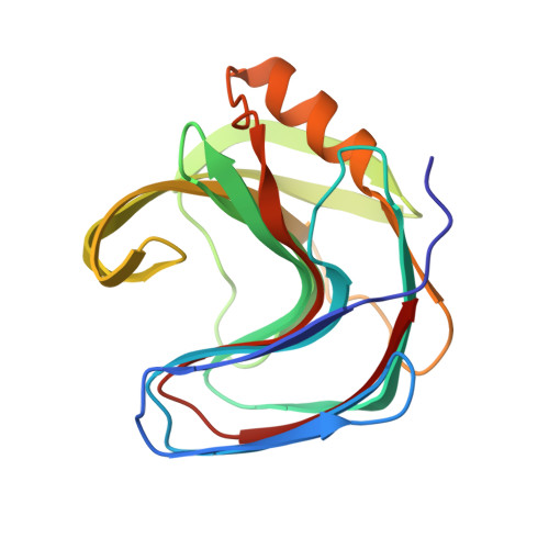
5TVV, 5TVY, 5TZO - PubMed Abstract:
We describe the computational design of proteins that bind the potent analgesic fentanyl. Our approach employs a fast docking algorithm to find shape complementary ligand placement in protein scaffolds, followed by design of the surrounding residues to optimize binding affinity. Co-crystal structures of the highest affinity binder reveal a highly preorganized binding site, and an overall architecture and ligand placement in close agreement with the design model. We use the designs to generate plant sensors for fentanyl by coupling ligand binding to design stability. The method should be generally useful for detecting toxic hydrophobic compounds in the environment.
- Department of Biochemistry, University of Washington, Seattle, United States.
Organizational Affiliation: