5TSZ | pdb_00005tsz

Crystal structure of Plasmodium vivax CelTOS


Experimental Data Snapshot

  • Method: X-RAY DIFFRACTION
  • Resolution: 3.00 Å
  • R-Value Free: 
    0.238 (Depositor), 0.243 (DCC) 
  • R-Value Work: 
    0.220 (Depositor), 0.223 (DCC) 
  • R-Value Observed: 
    0.221 (Depositor) 

wwPDB Validation 3D Report Full Report

Validation slider image for 5TSZ

This is version 1.1 of the entry. See complete history

Literature

Malaria parasite CelTOS targets the inner leaflet of cell membranes for pore-dependent disruption.

Jimah, J.R.Salinas, N.D.Sala-Rabanal, M.Jones, N.G.Sibley, L.D.Nichols, C.G.Schlesinger, P.H.Tolia, N.H.

(2016) Elife 5

  • DOI: https://doi.org/10.7554/eLife.20621
  • Primary Citation Related Structures: 
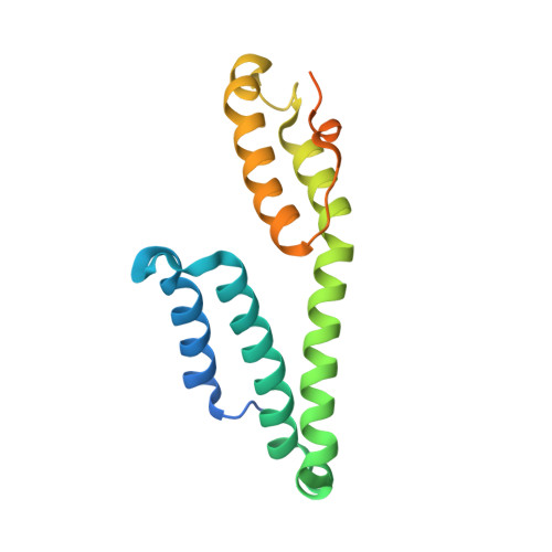
    5TSZ

  • PubMed Abstract: 

    Apicomplexan parasites contain a conserved protein CelTOS that, in malaria parasites, is essential for traversal of cells within the mammalian host and arthropod vector. However, the molecular role of CelTOS is unknown because it lacks sequence similarity to proteins of known function. Here, we determined the crystal structure of CelTOS and discovered CelTOS resembles proteins that bind to and disrupt membranes. In contrast to known membrane disruptors, CelTOS has a distinct architecture, specifically binds phosphatidic acid commonly present within the inner leaflet of plasma membranes, and potently disrupts liposomes composed of phosphatidic acid by forming pores. Microinjection of CelTOS into cells resulted in observable membrane damage. Therefore, CelTOS is unique as it achieves nearly universal inner leaflet cellular activity to enable the exit of parasites from cells during traversal. By providing novel molecular insight into cell traversal by apicomplexan parasites, our work facilitates the design of therapeutics against global pathogens.


  • Organizational Affiliation
    • Department of Molecular Microbiology, Washington University School of Medicine, Saint Louis, United States.

Macromolecule Content 

  • Total Structure Weight: 56.13 kDa 
  • Atom Count: 2,991 
  • Modeled Residue Count: 390 
  • Deposited Residue Count: 513 
  • Unique protein chains: 1

Macromolecules

Find similar proteins by:|  3D Structure
Entity ID: 1
MoleculeChains  Sequence LengthOrganismDetailsImage
Pv cell-traversal protein
A, B, C
171Plasmodium vivaxMutation(s): 0 
Gene Names: pvCelTOS
UniProt
Find proteins for Q53UB7 (Plasmodium vivax)
Explore Q53UB7 
Go to UniProtKB:  Q53UB7
Entity Groups
Sequence Clusters30% Identity50% Identity70% Identity90% Identity95% Identity100% Identity
UniProt GroupQ53UB7
Sequence Annotations
Expand
Reference Sequence

Experimental Data & Validation

Experimental Data

  • Method: X-RAY DIFFRACTION
  • Resolution: 3.00 Å
  • R-Value Free:  0.238 (Depositor), 0.243 (DCC) 
  • R-Value Work:  0.220 (Depositor), 0.223 (DCC) 
  • R-Value Observed: 0.221 (Depositor) 
Space Group: P 31 2 1
Unit Cell:
Length ( Å )Angle ( ˚ )
a = 158.96α = 90
b = 158.96β = 90
c = 64.09γ = 120
Software Package:
Software NamePurpose
PHENIXrefinement
XDSdata reduction
XDSdata scaling
SHARPphasing

Structure Validation

View Full Validation Report


Entry History 

Deposition Data

Revision History  (Full details and data files)

  • Version 1.0: 2016-12-28
    Type: Initial release
  • Version 1.1: 2024-03-06
    Changes: Data collection, Database references, Derived calculations