5TPX | pdb_00005tpx

Bromodomain from Plasmodium Faciparum Gcn5, complexed with compound


Experimental Data Snapshot

  • Method: X-RAY DIFFRACTION
  • Resolution: 2.10 Å
  • R-Value Free: 
    0.236 (Depositor), 0.196 (DCC) 
  • R-Value Work: 
    0.185 (Depositor) 
  • R-Value Observed: 
    0.188 (Depositor) 

Starting Model: experimental
View more details

wwPDB Validation 3D Report Full Report

Validation slider image for 5TPX

Ligand Structure Quality Assessment 


This is version 1.4 of the entry. See complete history

Literature

Discovery of a PCAF Bromodomain Chemical Probe.

Moustakim, M.Clark, P.G.Trulli, L.Fuentes de Arriba, A.L.Ehebauer, M.T.Chaikuad, A.Murphy, E.J.Mendez-Johnson, J.Daniels, D.Hou, C.D.Lin, Y.H.Walker, J.R.Hui, R.Yang, H.Dorrell, L.Rogers, C.M.Monteiro, O.P.Fedorov, O.Huber, K.V.Knapp, S.Heer, J.Dixon, D.J.Brennan, P.E.

(2017) Angew Chem Int Ed Engl 56: 827-831

  • DOI: https://doi.org/10.1002/anie.201610816
  • Primary Citation Related Structures: 
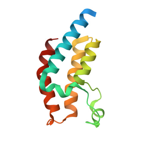
    5TPX

  • PubMed Abstract: 

    The p300/CBP-associated factor (PCAF) and related GCN5 bromodomain-containing lysine acetyl transferases are members of subfamily I of the bromodomain phylogenetic tree. Iterative cycles of rational inhibitor design and biophysical characterization led to the discovery of the triazolopthalazine-based L-45 (dubbed L-Moses) as the first potent, selective, and cell-active PCAF bromodomain (Brd) inhibitor. Synthesis from readily available (1R,2S)-(-)-norephedrine furnished L-45 in enantiopure form. L-45 was shown to disrupt PCAF-Brd histone H3.3 interaction in cells using a nanoBRET assay, and a co-crystal structure of L-45 with the homologous Brd PfGCN5 from Plasmodium falciparum rationalizes the high selectivity for PCAF and GCN5 bromodomains. Compound L-45 shows no observable cytotoxicity in peripheral blood mononuclear cells (PBMC), good cell-permeability, and metabolic stability in human and mouse liver microsomes, supporting its potential for in vivo use.


  • Organizational Affiliation
    • Structural Genomics Consortium & Target Discovery Institute, University of Oxford, NDM Research Building, Roosevelt Drive, Oxford, OX3 7DQ and OX3 7FZ, UK.

Macromolecule Content 

  • Total Structure Weight: 15.45 kDa 
  • Atom Count: 996 
  • Modeled Residue Count: 106 
  • Deposited Residue Count: 123 
  • Unique protein chains: 1

Macromolecules

Find similar proteins by:|  3D Structure
Entity ID: 1
MoleculeChains  Sequence LengthOrganismDetailsImage
Histone acetyltransferase GCN5123Plasmodium falciparum 3D7Mutation(s): 0 
Gene Names: PF3D7_0823300
EC: 2.3.1.48
UniProt
Find proteins for Q8IB67 (Plasmodium falciparum (isolate 3D7))
Explore Q8IB67 
Go to UniProtKB:  Q8IB67
Entity Groups
Sequence Clusters30% Identity50% Identity70% Identity90% Identity95% Identity100% Identity
UniProt GroupQ8IB67
Sequence Annotations
Expand
Reference Sequence

Experimental Data & Validation

Experimental Data

  • Method: X-RAY DIFFRACTION
  • Resolution: 2.10 Å
  • R-Value Free:  0.236 (Depositor), 0.196 (DCC) 
  • R-Value Work:  0.185 (Depositor) 
  • R-Value Observed: 0.188 (Depositor) 
Space Group: P 4 21 2
Unit Cell:
Length ( Å )Angle ( ˚ )
a = 75α = 90
b = 75β = 90
c = 49.62γ = 90
Software Package:
Software NamePurpose
HKL-2000data collection
HKL-2000data scaling
BUSTER-TNTrefinement
PDB_EXTRACTdata extraction
PHASERphasing
DENZOdata reduction
SCALEPACKdata scaling
BUSTERrefinement

Structure Validation

View Full Validation Report


Ligand Structure Quality Assessment 


Entry History 

Revision History  (Full details and data files)

  • Version 1.0: 2017-01-04
    Type: Initial release
  • Version 1.1: 2017-01-18
    Changes: Database references
  • Version 1.2: 2017-11-22
    Changes: Refinement description
  • Version 1.3: 2018-01-24
    Changes: Structure summary
  • Version 1.4: 2023-10-04
    Changes: Data collection, Database references, Refinement description