5TO3

Crystal structure of thrombin mutant W215A/E217A fused to EGF456 of thrombomodulin via a 31-residue linker and bound to PPACK


Experimental Data Snapshot

  • Method: X-RAY DIFFRACTION
  • Resolution: 2.34 Å
  • R-Value Free: 0.255 
  • R-Value Work: 0.214 
  • R-Value Observed: 0.216 

wwPDB Validation   3D Report Full Report


Ligand Structure Quality Assessment 


This is version 2.1 of the entry. See complete history


Literature

Rational Design of Protein C Activators.

Barranco-Medina, S.Murphy, M.Pelc, L.Chen, Z.Di Cera, E.Pozzi, N.

(2017) Sci Rep 7: 44596-44596

  • DOI: https://doi.org/10.1038/srep44596
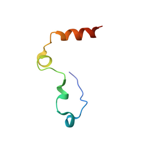
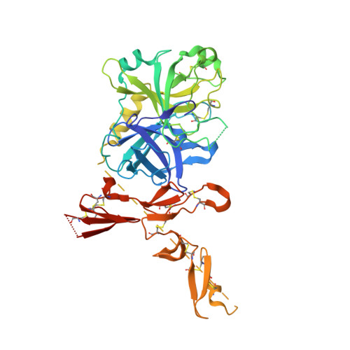
  • Primary Citation of Related Structures:  
    5TO3

  • PubMed Abstract: 

    In addition to its procoagulant and proinflammatory functions mediated by cleavage of fibrinogen and PAR1, the trypsin-like protease thrombin activates the anticoagulant protein C in a reaction that requires the cofactor thrombomodulin and the endothelial protein C receptor. Once in the circulation, activated protein C functions as an anticoagulant, anti-inflammatory and regenerative factor. Hence, availability of a protein C activator would afford a therapeutic for patients suffering from thrombotic disorders and a diagnostic tool for monitoring the level of protein C in plasma. Here, we present a fusion protein where thrombin and the EGF456 domain of thrombomodulin are connected through a peptide linker. The fusion protein recapitulates the functional and structural properties of the thrombin-thrombomodulin complex, prolongs the clotting time by generating pharmacological quantities of activated protein C and effectively diagnoses protein C deficiency in human plasma. Notably, these functions do not require exogenous thrombomodulin, unlike other anticoagulant thrombin derivatives engineered to date. These features make the fusion protein an innovative step toward the development of protein C activators of clinical and diagnostic relevance.


  • Organizational Affiliation

    Edward A. Doisy Department of Biochemistry and Molecular Biology, Saint Louis University School of Medicine, St. Louis, MO 63104 USA.


Macromolecules
Find similar proteins by:  (by identity cutoff)  |  3D Structure
Entity ID: 1
MoleculeChains Sequence LengthOrganismDetailsImage
Prothrombin46Homo sapiensMutation(s): 0 
Gene Names: F2
EC: 3.4.21.5
UniProt & NIH Common Fund Data Resources
Find proteins for P00734 (Homo sapiens)
Explore P00734 
Go to UniProtKB:  P00734
PHAROS:  P00734
GTEx:  ENSG00000180210 
Entity Groups  
Sequence Clusters30% Identity50% Identity70% Identity90% Identity95% Identity100% Identity
UniProt GroupP00734
Sequence Annotations
Expand
  • Reference Sequence
Find similar proteins by:  (by identity cutoff)  |  3D Structure
Entity ID: 2
MoleculeChains Sequence LengthOrganismDetailsImage
Prothrombin,Thrombomodulin409Homo sapiensMutation(s): 2 
Gene Names: F2THBDTHRM
EC: 3.4.21.5
UniProt & NIH Common Fund Data Resources
Find proteins for P00734 (Homo sapiens)
Explore P00734 
Go to UniProtKB:  P00734
PHAROS:  P00734
GTEx:  ENSG00000180210 
Find proteins for P07204 (Homo sapiens)
Explore P07204 
Go to UniProtKB:  P07204
PHAROS:  P07204
GTEx:  ENSG00000178726 
Entity Groups  
Sequence Clusters30% Identity50% Identity70% Identity90% Identity95% Identity100% Identity
UniProt GroupsP00734P07204
Sequence Annotations
Expand
  • Reference Sequence
Oligosaccharides

Help

Entity ID: 3
MoleculeChains Length2D Diagram Glycosylation3D Interactions
2-acetamido-2-deoxy-beta-D-glucopyranose-(1-4)-beta-D-mannopyranose-(1-4)-beta-D-mannopyranose-(1-4)-alpha-D-mannopyranose-(1-4)-[beta-D-mannopyranose-(1-6)]beta-D-mannopyranose-(1-4)-2-acetamido-2-deoxy-beta-D-glucopyranose
C
7N-Glycosylation
Glycosylation Resources
GlyTouCan:  G20640NX
GlyCosmos:  G20640NX
GlyGen:  G20640NX
Biologically Interesting Molecules (External Reference) 1 Unique
Experimental Data & Validation

Experimental Data

  • Method: X-RAY DIFFRACTION
  • Resolution: 2.34 Å
  • R-Value Free: 0.255 
  • R-Value Work: 0.214 
  • R-Value Observed: 0.216 
  • Space Group: C 2 2 21
Unit Cell:
Length ( Å )Angle ( ˚ )
a = 62.939α = 90
b = 162.174β = 90
c = 128.859γ = 90
Software Package:
Software NamePurpose
REFMACrefinement
HKL-2000data reduction
HKL-2000data scaling

Structure Validation

View Full Validation Report


Ligand Structure Quality Assessment 


Entry History & Funding Information

Deposition Data


Funding OrganizationLocationGrant Number
National Institutes of Health/National Heart, Lung, and Blood Institute (NIH/NHLBI)United States--

Revision History  (Full details and data files)

  • Version 1.0: 2017-03-29
    Type: Initial release
  • Version 1.1: 2017-09-06
    Changes: Author supporting evidence
  • Version 1.2: 2019-12-04
    Changes: Author supporting evidence
  • Version 2.0: 2020-07-29
    Type: Remediation
    Reason: Carbohydrate remediation
    Changes: Atomic model, Data collection, Derived calculations, Structure summary
  • Version 2.1: 2023-10-04
    Changes: Data collection, Database references, Derived calculations, Refinement description, Structure summary