5TLB

Scabin toxin from Streptomyces scabies in complex with NADH


Experimental Data Snapshot

  • Method: X-RAY DIFFRACTION
  • Resolution: 1.70 Å
  • R-Value Free: 0.196 
  • R-Value Work: 0.159 

wwPDB Validation   3D Report Full Report


Ligand Structure Quality Assessment 


This is version 1.3 of the entry. See complete history


Literature

Characterization of the catalytic signature of Scabin toxin, a DNA-targeting ADP-ribosyltransferase.

Lyons, B.Lugo, M.R.Carlin, S.Lidster, T.Merrill, A.R.

(2018) Biochem J 475: 225-245

  • DOI: https://doi.org/10.1042/BCJ20170818
  • Primary Citation of Related Structures:  
    5TLB, 5UVQ, 6APY

  • PubMed Abstract: 

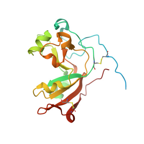
    Scabin was previously identified as a novel DNA-targeting mono-ADP-ribosyltransferase (mART) toxin from the plant pathogen 87.22 strain of Streptomyces scabies Scabin is a member of the Pierisin-like subgroup of mART toxins, since it targets DNA. An in-depth characterization of both the glycohydrolase and transferase enzymatic activities of Scabin was conducted. Several protein variants were developed based on an initial Scabin·DNA molecular model. Consequently, three residues were deemed important for DNA-binding and transferase activity. Trp128 and Trp155 are important for binding the DNA substrate and participate in the reaction mechanism, whereas Tyr129 was shown to be important only for DNA binding, but was not involved in the reaction mechanism. Trp128 and Trp155 are both conserved within the Pierisin-like toxins, whereas Tyr129 is a unique substitution within the group. Scabin showed substrate specificity toward double-stranded DNA containing a single-base overhang, as a model for single-stranded nicked DNA. The crystal structure of Scabin bound to NADH - a competitive inhibitor of Scabin - was determined, providing important insights into the active-site structure and Michaelis-Menten complex of the enzyme. Based on these results, a novel DNA-binding motif is proposed for Scabin with substrate and the key residues that may participate in the Scabin·NAD(+) complex are highlighted.


  • Organizational Affiliation

    Department of Molecular and Cellular Biology, University of Guelph, Guelph, Ontario, Canada N1G 2W1.


Macromolecules
Find similar proteins by:  (by identity cutoff)  |  3D Structure
Entity ID: 1
MoleculeChains Sequence LengthOrganismDetailsImage
Scabin200Streptomyces scabiei 87.22Mutation(s): 0 
Gene Names: SCAB_27771
EC: 2.4.2.31
UniProt
Find proteins for C9Z6T8 (Streptomyces scabiei (strain 87.22))
Explore C9Z6T8 
Go to UniProtKB:  C9Z6T8
Entity Groups  
Sequence Clusters30% Identity50% Identity70% Identity90% Identity95% Identity100% Identity
UniProt GroupC9Z6T8
Sequence Annotations
Expand
  • Reference Sequence
Small Molecules
Ligands 1 Unique
IDChains Name / Formula / InChI Key2D Diagram3D Interactions
NAI
Query on NAI

Download Ideal Coordinates CCD File 
B [auth A]1,4-DIHYDRONICOTINAMIDE ADENINE DINUCLEOTIDE
C21 H29 N7 O14 P2
BOPGDPNILDQYTO-NNYOXOHSSA-N
Binding Affinity Annotations 
IDSourceBinding Affinity
NAI Binding MOAD:  5TLB Kd: 7.00e+4 (nM) from 1 assay(s)
Experimental Data & Validation

Experimental Data

  • Method: X-RAY DIFFRACTION
  • Resolution: 1.70 Å
  • R-Value Free: 0.196 
  • R-Value Work: 0.159 
  • Space Group: C 1 2 1
Unit Cell:
Length ( Å )Angle ( ˚ )
a = 86.04α = 90
b = 62.26β = 100.3
c = 38.08γ = 90
Software Package:
Software NamePurpose
PHENIXrefinement
XDSdata reduction
XSCALEdata scaling
PHASERphasing

Structure Validation

View Full Validation Report


Ligand Structure Quality Assessment 


Entry History 

Deposition Data

Revision History  (Full details and data files)

  • Version 1.0: 2017-10-11
    Type: Initial release
  • Version 1.1: 2017-12-20
    Changes: Database references, Structure summary
  • Version 1.2: 2018-01-17
    Changes: Database references
  • Version 1.3: 2023-10-04
    Changes: Data collection, Database references, Refinement description