5TE2 | pdb_00005te2

Crystal structure of 7,8-diaminopelargonic acid synthase (BioA) from Mycobacterium tuberculosis, complexed with a mechanism-based inhibitor


Experimental Data Snapshot

  • Method: X-RAY DIFFRACTION
  • Resolution: 1.80 Å
  • R-Value Free: 
    0.193 (Depositor), 0.195 (DCC) 
  • R-Value Work: 
    0.168 (Depositor), 0.170 (DCC) 
  • R-Value Observed: 
    0.169 (Depositor) 

Starting Model: experimental
View more details

wwPDB Validation 3D Report Full Report

Validation slider image for 5TE2

Ligand Structure Quality Assessment 


This is version 1.4 of the entry. See complete history

Literature

Rational Optimization of Mechanism-Based Inhibitors through Determination of the Microscopic Rate Constants of Inactivation.

Eiden, C.G.Maize, K.M.Finzel, B.C.Lipscomb, J.D.Aldrich, C.C.

(2017) J Am Chem Soc 139: 7132-7135

  • DOI: https://doi.org/10.1021/jacs.7b00962
  • Primary Citation Related Structures: 
    5TE2

  • PubMed Abstract: 

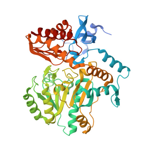
    Mechanism-based inhibitors (MBIs) are widely employed in chemistry, biology, and medicine because of their exquisite specificity and sustained duration of inhibition. Optimization of MBIs is complicated because of time-dependent inhibition resulting from multistep inactivation mechanisms. The global kinetic parameters k inact and K I have been used to characterize MBIs, but they provide far less information than is commonly assumed, as shown by derivation and simulation of these parameters. We illustrate an alternative and more rigorous approach for MBI characterization through determination of the individual microscopic rate constants. Kinetic analysis revealed the rate-limiting step of inactivation of the PLP-dependent enzyme BioA by dihydro-(1,4)-pyridone 1. This knowledge was subsequently applied to rationally design a second-generation inhibitor scaffold with a nearly optimal maximum inactivation rate (0.48 min -1 ).


  • Organizational Affiliation
    • Department of Medicinal Chemistry, University of Minnesota , Minneapolis, Minnesota 55455, United States.

Macromolecule Content 

  • Total Structure Weight: 98.19 kDa 
  • Atom Count: 6,914 
  • Modeled Residue Count: 856 
  • Deposited Residue Count: 914 
  • Unique protein chains: 1

Macromolecules

Find similar proteins by:|  3D Structure
Entity ID: 1
MoleculeChains  Sequence LengthOrganismDetailsImage
Adenosylmethionine-8-amino-7-oxononanoate aminotransferaseA [auth B],
B [auth A]
457Mycobacterium tuberculosis variant bovis AF2122/97Mutation(s): 0 
Gene Names: bioAMb1595
EC: 2.6.1.62
UniProt
Find proteins for P0A4X7 (Mycobacterium bovis (strain ATCC BAA-935 / AF2122/97))
Explore P0A4X7 
Go to UniProtKB:  P0A4X7
Entity Groups
Sequence Clusters30% Identity50% Identity70% Identity90% Identity95% Identity100% Identity
UniProt GroupP0A4X7
Sequence Annotations
Expand
Reference Sequence

Small Molecules

Ligands 4 Unique
IDChains Name / Formula / InChI Key2D Diagram3D Interactions
7B9

Query on 7B9


Download:Ideal Coordinates CCD File
C [auth B],
F [auth A]
[5-hydroxy-4-({[6-(3-hydroxypropyl)-4-oxo-1,4-dihydropyridin-3-yl]amino}methyl)-6-methylpyridin-3-yl]methyl dihydrogen phosphate
C16 H22 N3 O7 P
LWVUNNHCQMOSIS-UHFFFAOYSA-N
PGE

Query on PGE


Download:Ideal Coordinates CCD File
G [auth A]TRIETHYLENE GLYCOL
C6 H14 O4
ZIBGPFATKBEMQZ-UHFFFAOYSA-N
PEG

Query on PEG


Download:Ideal Coordinates CCD File
D [auth B]DI(HYDROXYETHYL)ETHER
C4 H10 O3
MTHSVFCYNBDYFN-UHFFFAOYSA-N
EDO

Query on EDO


Download:Ideal Coordinates CCD File
E [auth B]1,2-ETHANEDIOL
C2 H6 O2
LYCAIKOWRPUZTN-UHFFFAOYSA-N

Experimental Data & Validation

Experimental Data

  • Method: X-RAY DIFFRACTION
  • Resolution: 1.80 Å
  • R-Value Free:  0.193 (Depositor), 0.195 (DCC) 
  • R-Value Work:  0.168 (Depositor), 0.170 (DCC) 
  • R-Value Observed: 0.169 (Depositor) 
Space Group: P 21 21 21
Unit Cell:
Length ( Å )Angle ( ˚ )
a = 62.696α = 90
b = 66.364β = 90
c = 203.956γ = 90
Software Package:
Software NamePurpose
PHENIXrefinement
Aimlessdata scaling
PHASERphasing
PDB_EXTRACTdata extraction
XDSdata reduction

Structure Validation

View Full Validation Report


Ligand Structure Quality Assessment 


Entry History 

& Funding Information

Deposition Data


Funding OrganizationLocationGrant Number
National Institutes of Health/National Institute Of Allergy and Infectious Diseases (NIH/NIAID)United StatesR01 AI091790

Revision History  (Full details and data files)

  • Version 1.0: 2017-05-31
    Type: Initial release
  • Version 1.1: 2017-06-14
    Changes: Database references
  • Version 1.2: 2017-09-20
    Changes: Author supporting evidence
  • Version 1.3: 2019-12-11
    Changes: Author supporting evidence
  • Version 1.4: 2023-10-04
    Changes: Data collection, Database references, Refinement description