5TC3

Structure of IMP dehydrogenase from Ashbya gossypii bound to ATP and GDP


Experimental Data Snapshot

  • Method: X-RAY DIFFRACTION
  • Resolution: 2.46 Å
  • R-Value Free: 0.242 
  • R-Value Work: 0.216 
  • R-Value Observed: 0.218 

wwPDB Validation   3D Report Full Report


Ligand Structure Quality Assessment 


This is version 1.1 of the entry. See complete history


Literature

A nucleotide-controlled conformational switch modulates the activity of eukaryotic IMP dehydrogenases.

Buey, R.M.Fernandez-Justel, D.Marcos-Alcalde, I.Winter, G.Gomez-Puertas, P.de Pereda, J.M.Luis Revuelta, J.

(2017) Sci Rep 7: 2648-2648

  • DOI: https://doi.org/10.1038/s41598-017-02805-x
  • Primary Citation of Related Structures:  
    5MCP, 5TC3

  • PubMed Abstract: 

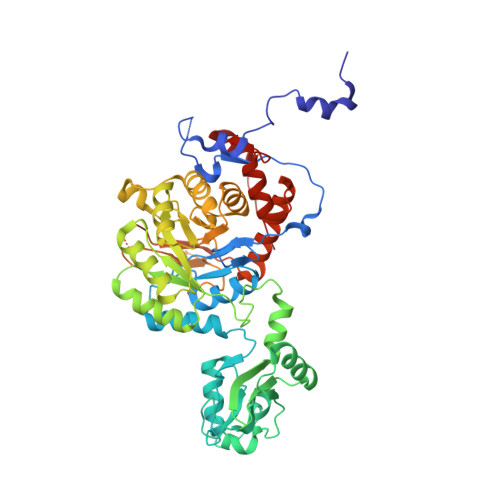
    Inosine-5'-monophosphate dehydrogenase (IMPDH) is an essential enzyme for nucleotide metabolism and cell proliferation. Despite IMPDH is the target of drugs with antiviral, immunosuppressive and antitumor activities, its physiological mechanisms of regulation remain largely unknown. Using the enzyme from the industrial fungus Ashbya gossypii, we demonstrate that the binding of adenine and guanine nucleotides to the canonical nucleotide binding sites of the regulatory Bateman domain induces different enzyme conformations with significantly distinct catalytic activities. Thereby, the comparison of their high-resolution structures defines the mechanistic and structural details of a nucleotide-controlled conformational switch that allosterically modulates the catalytic activity of eukaryotic IMPDHs. Remarkably, retinopathy-associated mutations lie within the mechanical hinges of the conformational change, highlighting its physiological relevance. Our results expand the mechanistic repertoire of Bateman domains and pave the road to new approaches targeting IMPDHs.


  • Organizational Affiliation

    Metabolic Engineering Group, Dpto. Microbiología y Genética, Universidad de Salamanca, Campus Miguel de Unamuno, 37007, Salamanca, Spain. ruben.martinez@usal.es.


Macromolecules
Find similar proteins by:  (by identity cutoff)  |  3D Structure
Entity ID: 1
MoleculeChains Sequence LengthOrganismDetailsImage
Inosine-5'-monophosphate dehydrogenase
A, B
523Eremothecium gossypii ATCC 10895Mutation(s): 0 
Gene Names: AGOS_AER117W
EC: 1.1.1.205
UniProt
Find proteins for Q756Z6 (Eremothecium gossypii (strain ATCC 10895 / CBS 109.51 / FGSC 9923 / NRRL Y-1056))
Explore Q756Z6 
Go to UniProtKB:  Q756Z6
Entity Groups  
Sequence Clusters30% Identity50% Identity70% Identity90% Identity95% Identity100% Identity
UniProt GroupQ756Z6
Sequence Annotations
Expand
  • Reference Sequence
Small Molecules
Ligands 4 Unique
IDChains Name / Formula / InChI Key2D Diagram3D Interactions
ATP
Query on ATP

Download Ideal Coordinates CCD File 
D [auth A],
I [auth B]
ADENOSINE-5'-TRIPHOSPHATE
C10 H16 N5 O13 P3
ZKHQWZAMYRWXGA-KQYNXXCUSA-N
GDP
Query on GDP

Download Ideal Coordinates CCD File 
E [auth A],
F [auth A],
J [auth B],
K [auth B]
GUANOSINE-5'-DIPHOSPHATE
C10 H15 N5 O11 P2
QGWNDRXFNXRZMB-UUOKFMHZSA-N
5GP
Query on 5GP

Download Ideal Coordinates CCD File 
C [auth A],
H [auth B]
GUANOSINE-5'-MONOPHOSPHATE
C10 H14 N5 O8 P
RQFCJASXJCIDSX-UUOKFMHZSA-N
ACT
Query on ACT

Download Ideal Coordinates CCD File 
G [auth A]ACETATE ION
C2 H3 O2
QTBSBXVTEAMEQO-UHFFFAOYSA-M
Experimental Data & Validation

Experimental Data

  • Method: X-RAY DIFFRACTION
  • Resolution: 2.46 Å
  • R-Value Free: 0.242 
  • R-Value Work: 0.216 
  • R-Value Observed: 0.218 
  • Space Group: I 4
Unit Cell:
Length ( Å )Angle ( ˚ )
a = 147.88α = 90
b = 147.88β = 90
c = 103.542γ = 90
Software Package:
Software NamePurpose
PHENIXrefinement
XDSdata reduction
XSCALEdata scaling
PHASERphasing

Structure Validation

View Full Validation Report


Ligand Structure Quality Assessment 


Entry History 

Deposition Data

Revision History  (Full details and data files)

  • Version 1.0: 2017-06-14
    Type: Initial release
  • Version 1.1: 2024-01-17
    Changes: Data collection, Database references, Refinement description