5T9J | pdb_00005t9j

Crystal Structure of human GEN1 in complex with Holliday junction DNA in the upper interface


Experimental Data Snapshot

  • Method: X-RAY DIFFRACTION
  • Resolution: 3.00 Å
  • R-Value Free: 
    0.241 (Depositor), 0.248 (DCC) 
  • R-Value Work: 
    0.199 (Depositor), 0.204 (DCC) 
  • R-Value Observed: 
    0.201 (Depositor) 

wwPDB Validation 3D Report Full Report

Validation slider image for 5T9J

This is version 1.1 of the entry. See complete history

Literature

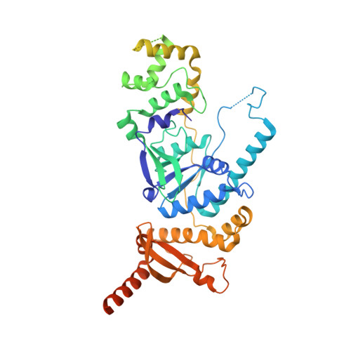
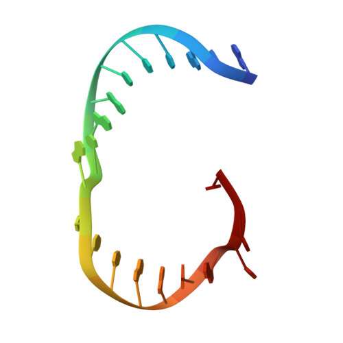
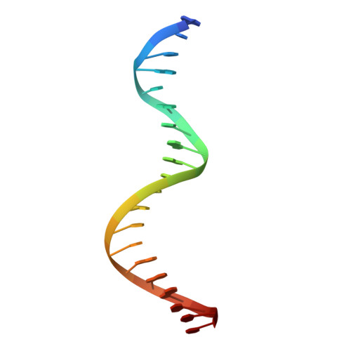
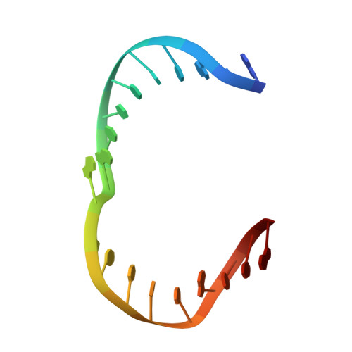
Human Holliday junction resolvase GEN1 uses a chromodomain for efficient DNA recognition and cleavage.

Lee, S.H.Princz, L.N.Klugel, M.F.Habermann, B.Pfander, B.Biertumpfel, C.

(2015) Elife 4

  • DOI: https://doi.org/10.7554/eLife.12256
  • Primary Citation Related Structures: 
    5T9J

  • PubMed Abstract: 

    Holliday junctions (HJs) are key DNA intermediates in homologous recombination. They link homologous DNA strands and have to be faithfully removed for proper DNA segregation and genome integrity. Here, we present the crystal structure of human HJ resolvase GEN1 complexed with DNA at 3.0 Å resolution. The GEN1 core is similar to other Rad2/XPG nucleases. However, unlike other members of the superfamily, GEN1 contains a chromodomain as an additional DNA interaction site. Chromodomains are known for their chromatin-targeting function in chromatin remodelers and histone(de)acetylases but they have not previously been found in nucleases. The GEN1 chromodomain directly contacts DNA and its truncation severely hampers GEN1's catalytic activity. Structure-guided mutations in vitro and in vivo in yeast validated our mechanistic findings. Our study provides the missing structure in the Rad2/XPG family and insights how a well-conserved nuclease core acquires versatility in recognizing diverse substrates for DNA repair and maintenance.


  • Organizational Affiliation
    • Department of Structural Cell Biology, Molecular Mechanisms of DNA Repair, Max Planck Institute of Biochemistry, Martinsried, Germany.

Macromolecule Content 

  • Total Structure Weight: 142.76 kDa 
  • Atom Count: 8,002 
  • Modeled Residue Count: 859 
  • Deposited Residue Count: 1,092 
  • Unique protein chains: 1
  • Unique nucleic acid chains: 4

Macromolecules


Find similar proteins by:|  3D Structure
Entity ID: 1
MoleculeChains  Sequence LengthOrganismDetailsImage
Flap endonuclease GEN homolog 1
A, B
506Homo sapiensMutation(s): 3 
Gene Names: GEN1
EC: 3.1
UniProt & NIH Common Fund Data Resources
Find proteins for Q17RS7 (Homo sapiens)
Explore Q17RS7 
Go to UniProtKB:  Q17RS7
PHAROS:  Q17RS7
GTEx:  ENSG00000178295 
Entity Groups
Sequence Clusters30% Identity50% Identity70% Identity90% Identity95% Identity100% Identity
UniProt GroupQ17RS7
Sequence Annotations
Expand
Reference Sequence
Find similar nucleic acids by:  Sequence
Entity ID: 2
MoleculeChains LengthOrganismImage
DNA (5'-D(*DGP*DAP*DAP*DTP*DTP*DCP*DCP*DGP*DGP*DAP*DTP*DTP*DAP*DGP*DGP*DGP*DAP*DTP*DGP*DC)-3')20synthetic construct
Sequence Annotations
Expand
Reference Sequence
Find similar nucleic acids by:  Sequence
Entity ID: 3
MoleculeChains LengthOrganismImage
DNA (5'-D(*DGP*DCP*DAP*DTP*DCP*DCP*DCP*DTP*DAP*DAP*DGP*DCP*DTP*DCP*DCP*DAP*DTP*DCP*DGP*DT)-3')20synthetic construct
Sequence Annotations
Expand
Reference Sequence
Find similar nucleic acids by:  Sequence
Entity ID: 4
MoleculeChains LengthOrganismImage
DNA (5'-D(*DAP*DCP*DGP*DAP*DTP*DGP*DGP*DAP*DGP*DCP*DCP*DGP*DCP*DTP*DAP*DGP*DGP*DCP*DTP*DC)-3')20synthetic construct
Sequence Annotations
Expand
Reference Sequence
Find similar nucleic acids by:  Sequence
Entity ID: 5
MoleculeChains LengthOrganismImage
DNA (5'-D(*DGP*DAP*DGP*DCP*DCP*DTP*DAP*DGP*DCP*DGP*DTP*DCP*DCP*DGP*DGP*DAP*DAP*DTP*DTP*DC)-3')20synthetic construct
Sequence Annotations
Expand
Reference Sequence

Small Molecules

Ligands 2 Unique
IDChains Name / Formula / InChI Key2D Diagram3D Interactions
GOL

Query on GOL


Download:Ideal Coordinates CCD File
G [auth A]
H [auth A]
I [auth A]
J [auth A]
K [auth A]
G [auth A],
H [auth A],
I [auth A],
J [auth A],
K [auth A],
L [auth A],
M [auth A],
O [auth B],
P [auth B],
Q [auth B],
R [auth B],
S [auth B],
U [auth C],
V [auth C],
W [auth C]
GLYCEROL
C3 H8 O3
PEDCQBHIVMGVHV-UHFFFAOYSA-N
MG

Query on MG


Download:Ideal Coordinates CCD File
N [auth A],
T [auth B]
MAGNESIUM ION
Mg
JLVVSXFLKOJNIY-UHFFFAOYSA-N

Experimental Data & Validation

Experimental Data

  • Method: X-RAY DIFFRACTION
  • Resolution: 3.00 Å
  • R-Value Free:  0.241 (Depositor), 0.248 (DCC) 
  • R-Value Work:  0.199 (Depositor), 0.204 (DCC) 
  • R-Value Observed: 0.201 (Depositor) 
Space Group: P 32
Unit Cell:
Length ( Å )Angle ( ˚ )
a = 86.94α = 90
b = 86.94β = 90
c = 200.724γ = 120
Software Package:
Software NamePurpose
autoSHARPphasing
PHENIXrefinement
PDB_EXTRACTdata extraction
XDSdata reduction
XDSdata scaling

Structure Validation

View Full Validation Report


Entry History 

& Funding Information

Deposition Data


Funding OrganizationLocationGrant Number
Max Planck SocietyGermanyMax Planck Research Group Leader Program

Revision History  (Full details and data files)

  • Version 1.0: 2016-09-21
    Type: Initial release
  • Version 1.1: 2024-05-08
    Changes: Data collection, Database references, Refinement description