Identification of Two Phosphate Starvation-induced Wall Teichoic Acid Hydrolases Provides First Insights into the Degradative Pathway of a Key Bacterial Cell Wall Component.
Myers, C.L., Li, F.K., Koo, B.M., El-Halfawy, O.M., French, S., Gross, C.A., Strynadka, N.C., Brown, E.D.(2016) J Biol Chem 291: 26066-26082
- PubMed: 27780866 Search on PubMedSearch on PubMed Central
- DOI: https://doi.org/10.1074/jbc.M116.760447
- Primary Citation Related Structures:
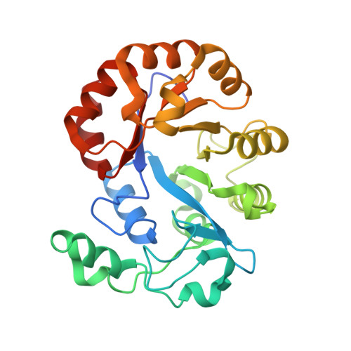
5T91, 5T9B, 5T9C - PubMed Abstract:
The cell wall of most Gram-positive bacteria contains equal amounts of peptidoglycan and the phosphate-rich glycopolymer wall teichoic acid (WTA). During phosphate-limited growth of the Gram-positive model organism Bacillus subtilis 168, WTA is lost from the cell wall in a response mediated by the PhoPR two-component system, which regulates genes involved in phosphate conservation and acquisition. It has been thought that WTA provides a phosphate source to sustain growth during starvation conditions; however, WTA degradative pathways have not been described for this or any condition of bacterial growth. Here, we uncover roles for the Bacillus subtilis PhoP regulon genes glpQ and phoD as encoding secreted phosphodiesterases that function in WTA metabolism during phosphate starvation. Unlike the parent 168 strain, ΔglpQ or ΔphoD mutants retained WTA and ceased growth upon phosphate limitation. Characterization of GlpQ and PhoD enzymatic activities, in addition to X-ray crystal structures of GlpQ, revealed distinct mechanisms of WTA depolymerization for the two enzymes; GlpQ catalyzes exolytic cleavage of individual monomer units, and PhoD catalyzes endo-hydrolysis at nonspecific sites throughout the polymer. The combination of these activities appears requisite for the utilization of WTA as a phosphate reserve. Phenotypic characterization of the ΔglpQ and ΔphoD mutants revealed altered cell morphologies and effects on autolytic activity and antibiotic susceptibilities that, unexpectedly, also occurred in phosphate-replete conditions. Our findings offer novel insight into the B. subtilis phosphate starvation response and implicate WTA hydrolase activity as a determinant of functional properties of the Gram-positive cell envelope.
- From the Department of Biochemistry and Biomedical Sciences and.
Organizational Affiliation: