5T5W | pdb_00005t5w

Structure of an affinity matured lambda-IFN/IFN-lambdaR1/IL-10Rbeta receptor complex


Experimental Data Snapshot

  • Method: X-RAY DIFFRACTION
  • Resolution: 2.85 Å
  • R-Value Free: 
    0.253 (Depositor), 0.250 (DCC) 
  • R-Value Work: 
    0.198 (Depositor), 0.200 (DCC) 
  • R-Value Observed: 
    0.201 (Depositor) 

Starting Models: experimental
View more details

wwPDB Validation   3D Report Full Report


Ligand Structure Quality Assessment 


This is version 1.9 of the entry. See complete history


Literature

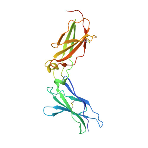
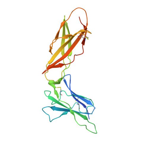
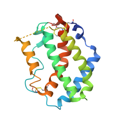
The IFN-lambda-IFN-lambda R1-IL-10R beta Complex Reveals Structural Features Underlying Type III IFN Functional Plasticity.

Mendoza, J.L.Schneider, W.M.Hoffmann, H.H.Vercauteren, K.Jude, K.M.Xiong, A.Moraga, I.Horton, T.M.Glenn, J.S.de Jong, Y.P.Rice, C.M.Garcia, K.C.

(2017) Immunity 46: 379-392

  • DOI: https://doi.org/10.1016/j.immuni.2017.02.017
  • Primary Citation of Related Structures:  
    5T5W

  • PubMed Abstract: 

    Type III interferons (IFN-λs) signal through a heterodimeric receptor complex composed of the IFN-λR1 subunit, specific for IFN-λs, and interleukin-10Rβ (IL-10Rβ), which is shared by multiple cytokines in the IL-10 superfamily. Low affinity of IL-10Rβ for cytokines has impeded efforts aimed at crystallizing cytokine-receptor complexes. We used yeast surface display to engineer a higher-affinity IFN-λ variant, H11, which enabled crystallization of the ternary complex. The structure revealed that IL-10Rβ uses a network of tyrosine residues as hydrophobic anchor points to engage IL-10 family cytokines that present complementary hydrophobic binding patches, explaining its role as both a cross-reactive but cytokine-specific receptor. H11 elicited increased anti-proliferative and antiviral activities in vitro and in vivo. In contrast, engineered higher-affinity type I IFNs did not increase antiviral potency over wild-type type I IFNs. Our findings provide insight into cytokine recognition by the IL-10R family and highlight the plasticity of type III interferon signaling and its therapeutic potential.


  • Organizational Affiliation
    • Howard Hughes Medical Institute, Department of Molecular and Cellular Physiology and Department of Structural Biology, Stanford University School of Medicine, Stanford, CA 94305, USA.

Macromolecules
Find similar proteins by:  (by identity cutoff)  |  3D Structure
Entity ID: 1
MoleculeChains Sequence LengthOrganismDetailsImage
Interleukin-10 receptor subunit beta214Homo sapiensMutation(s): 5 
Gene Names: IL10RBCRFB4D21S58D21S66
UniProt & NIH Common Fund Data Resources
Find proteins for Q08334 (Homo sapiens)
Explore Q08334 
Go to UniProtKB:  Q08334
PHAROS:  Q08334
GTEx:  ENSG00000243646 
Entity Groups  
Sequence Clusters30% Identity50% Identity70% Identity90% Identity95% Identity100% Identity
UniProt GroupQ08334
Sequence Annotations
Expand
  • Reference Sequence
Find similar proteins by:  (by identity cutoff)  |  3D Structure
Entity ID: 2
MoleculeChains Sequence LengthOrganismDetailsImage
Interferon lambda receptor 1218Homo sapiensMutation(s): 0 
Gene Names: IFNLR1IL28RALICR2
UniProt & NIH Common Fund Data Resources
Find proteins for Q8IU57 (Homo sapiens)
Explore Q8IU57 
Go to UniProtKB:  Q8IU57
PHAROS:  Q8IU57
GTEx:  ENSG00000185436 
Entity Groups  
Sequence Clusters30% Identity50% Identity70% Identity90% Identity95% Identity100% Identity
UniProt GroupQ8IU57
Glycosylation
Glycosylation Sites: 3Go to GlyGen: Q8IU57-1
Sequence Annotations
Expand
  • Reference Sequence
Find similar proteins by:  (by identity cutoff)  |  3D Structure
Entity ID: 3
MoleculeChains Sequence LengthOrganismDetailsImage
Interferon lambda-3176Homo sapiensMutation(s): 4 
Gene Names: IFNL3IL28BIL28CZCYTO22
UniProt & NIH Common Fund Data Resources
Find proteins for Q8IZI9 (Homo sapiens)
Explore Q8IZI9 
Go to UniProtKB:  Q8IZI9
PHAROS:  Q8IZI9
GTEx:  ENSG00000197110 
Entity Groups  
Sequence Clusters30% Identity50% Identity70% Identity90% Identity95% Identity100% Identity
UniProt GroupQ8IZI9
Sequence Annotations
Expand
  • Reference Sequence
Experimental Data & Validation

Experimental Data

  • Method: X-RAY DIFFRACTION
  • Resolution: 2.85 Å
  • R-Value Free:  0.253 (Depositor), 0.250 (DCC) 
  • R-Value Work:  0.198 (Depositor), 0.200 (DCC) 
  • R-Value Observed: 0.201 (Depositor) 
Space Group: P 31 2 1
Unit Cell:
Length ( Å )Angle ( ˚ )
a = 106.79α = 90
b = 106.79β = 90
c = 129.71γ = 120
Software Package:
Software NamePurpose
PHENIXrefinement
XSCALEdata scaling
PDB_EXTRACTdata extraction
XDSdata reduction
PHASERphasing

Structure Validation

View Full Validation Report


Ligand Structure Quality Assessment 


Entry History & Funding Information

Deposition Data


Funding OrganizationLocationGrant Number
National Institutes of Health/National Institute Of Allergy and Infectious Diseases (NIH/NIAID)United States1U19AI109662

Revision History  (Full details and data files)

  • Version 1.0: 2017-03-29
    Type: Initial release
  • Version 1.1: 2017-04-05
    Changes: Database references
  • Version 1.2: 2017-08-09
    Changes: Database references
  • Version 1.3: 2017-09-13
    Changes: Author supporting evidence
  • Version 1.4: 2018-08-15
    Changes: Data collection, Database references
  • Version 1.5: 2019-12-11
    Changes: Author supporting evidence
  • Version 1.6: 2020-07-29
    Type: Remediation
    Reason: Carbohydrate remediation
    Changes: Data collection, Derived calculations, Structure summary
  • Version 1.7: 2021-03-24
    Changes: Source and taxonomy, Structure summary
  • Version 1.8: 2023-10-04
    Changes: Data collection, Database references, Refinement description
  • Version 1.9: 2024-10-23
    Changes: Structure summary