5T4I

A Novel domain in human EXOG converts apoptotic endonuclease to DNA-repair enzyme


Experimental Data Snapshot

  • Method: X-RAY DIFFRACTION
  • Resolution: 2.39 Å
  • R-Value Free: 0.229 
  • R-Value Work: 0.191 
  • R-Value Observed: 0.193 

wwPDB Validation   3D Report Full Report


This is version 1.1 of the entry. See complete history


Literature

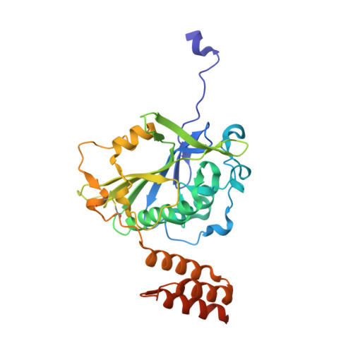
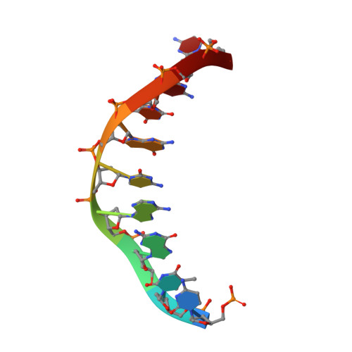
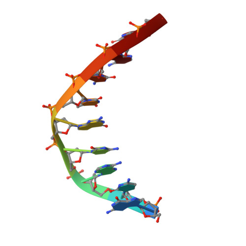
A domain in human EXOG converts apoptotic endonuclease to DNA-repair exonuclease.

Szymanski, M.R.Yu, W.Gmyrek, A.M.White, M.A.Molineux, I.J.Lee, J.C.Yin, Y.W.

(2017) Nat Commun 8: 14959-14959

  • DOI: https://doi.org/10.1038/ncomms14959
  • Primary Citation of Related Structures:  
    5T3V, 5T40, 5T4I, 5T5C

  • PubMed Abstract: 

    Human EXOG (hEXOG) is a 5'-exonuclease that is crucial for mitochondrial DNA repair; the enzyme belongs to a nonspecific nuclease family that includes the apoptotic endonuclease EndoG. Here we report biochemical and structural studies of hEXOG, including structures in its apo form and in a complex with DNA at 1.81 and 1.85 Å resolution, respectively. A Wing domain, absent in other ββα-Me members, suppresses endonuclease activity, but confers on hEXOG a strong 5'-dsDNA exonuclease activity that precisely excises a dinucleotide using an intrinsic 'tape-measure'. The symmetrical apo hEXOG homodimer becomes asymmetrical upon binding to DNA, providing a structural basis for how substrate DNA bound to one active site allosterically regulates the activity of the other. These properties of hEXOG suggest a pathway for mitochondrial BER that provides an optimal substrate for subsequent gap-filling synthesis by DNA polymerase γ.


  • Organizational Affiliation

    Department of Pharmacology and Toxicology, University of Texas Medical Branch, Galveston, Texas 77555, USA.


Macromolecules

Find similar proteins by:  (by identity cutoff)  |  3D Structure
Entity ID: 1
MoleculeChains Sequence LengthOrganismDetailsImage
Nuclease EXOG, mitochondrial
A, B
317Homo sapiensMutation(s): 1 
Gene Names: EXOGENDOGL1ENDOGL2ENGL
EC: 3.1.30
UniProt & NIH Common Fund Data Resources
Find proteins for Q9Y2C4 (Homo sapiens)
Explore Q9Y2C4 
Go to UniProtKB:  Q9Y2C4
PHAROS:  Q9Y2C4
GTEx:  ENSG00000157036 
Entity Groups  
Sequence Clusters30% Identity50% Identity70% Identity90% Identity95% Identity100% Identity
UniProt GroupQ9Y2C4
Sequence Annotations
Expand
  • Reference Sequence

Find similar nucleic acids by:  Sequence   |   3D Structure  

Entity ID: 2
MoleculeChains LengthOrganismImage
DNA (5'-D(P*CP*TP*GP*AP*CP*GP*TP*GP*C)-3')
C, E
9DNA launch vector pDE-GFP2
Sequence Annotations
Expand
  • Reference Sequence

Find similar nucleic acids by:  Sequence   |   3D Structure  

Entity ID: 3
MoleculeChains LengthOrganismImage
DNA (5'-D(*GP*CP*AP*CP*GP*TP*CP*AP*G)-3')
D, F
9DNA launch vector pDE-GFP2
Sequence Annotations
Expand
  • Reference Sequence
Experimental Data & Validation

Experimental Data

  • Method: X-RAY DIFFRACTION
  • Resolution: 2.39 Å
  • R-Value Free: 0.229 
  • R-Value Work: 0.191 
  • R-Value Observed: 0.193 
  • Space Group: P 21 21 21
Unit Cell:
Length ( Å )Angle ( ˚ )
a = 73.508α = 90
b = 79.723β = 90
c = 138.87γ = 90
Software Package:
Software NamePurpose
HKL-2000data collection
HKL-2000data scaling
PHENIXrefinement
PDB_EXTRACTdata extraction
HKL-2000data reduction
PHENIXphasing

Structure Validation

View Full Validation Report


Entry History 

Deposition Data

Revision History  (Full details and data files)

  • Version 1.0: 2017-05-17
    Type: Initial release
  • Version 1.1: 2023-10-04
    Changes: Data collection, Database references, Derived calculations, Refinement description