Structure of the Ebola virus envelope protein MPER/TM domain and its interaction with the fusion loop explains their fusion activity.
Lee, J., Nyenhuis, D.A., Nelson, E.A., Cafiso, D.S., White, J.M., Tamm, L.K.(2017) Proc Natl Acad Sci U S A 114: E7987-E7996
- PubMed: 28874543 Search on PubMedSearch on PubMed Central
- DOI: https://doi.org/10.1073/pnas.1708052114
- Primary Citation Related Structures:
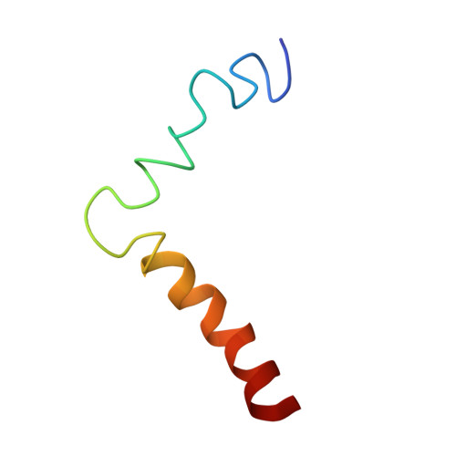
5T42 - PubMed Abstract:
Ebolavirus (EBOV), an enveloped filamentous RNA virus causing severe hemorrhagic fever, enters cells by macropinocytosis and membrane fusion in a late endosomal compartment. Fusion is mediated by the EBOV envelope glycoprotein GP, which consists of subunits GP1 and GP2. GP1 binds to cellular receptors, including Niemann-Pick C1 (NPC1) protein, and GP2 is responsible for low pH-induced membrane fusion. Proteolytic cleavage and NPC1 binding at endosomal pH lead to conformational rearrangements of GP2 that include exposing the hydrophobic fusion loop (FL) for insertion into the cellular target membrane and forming a six-helix bundle structure. Although major portions of the GP2 structure have been solved in pre- and postfusion states and although current models place the transmembrane (TM) and FL domains of GP2 in close proximity at critical steps of membrane fusion, their structures in membrane environments, and especially interactions between them, have not yet been characterized. Here, we present the structure of the membrane proximal external region (MPER) connected to the TM domain: i.e., the missing parts of the EBOV GP2 structure. The structure, solved by solution NMR and EPR spectroscopy in membrane-mimetic environments, consists of a helix-turn-helix architecture that is independent of pH. Moreover, the MPER region is shown to interact in the membrane interface with the previously determined structure of the EBOV FL through several critical aromatic residues. Mutation of aromatic and neighboring residues in both binding partners decreases fusion and viral entry, highlighting the functional importance of the MPER/TM-FL interaction in EBOV entry and fusion.
- Center for Membrane and Cell Physiology, University of Virginia, Charlottesville, VA 22908.
Organizational Affiliation: