


Thaumatin Structure at pH 6.0
Masuda, T., Sano, A., Murata, K., Okubo, K., Suzuki, M., Mikami, B.To be published.
Experimental Data Snapshot
Starting Model: experimental
View more details
Entity ID: 1 | |||||
|---|---|---|---|---|---|
| Molecule | Chains | Sequence Length | Organism | Details | Image |
| Thaumatin I | 207 | Thaumatococcus daniellii | Mutation(s): 0 | 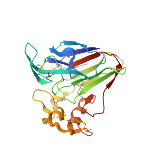 | |
UniProt | |||||
Entity Groups | |||||
| Sequence Clusters | 30% Identity50% Identity70% Identity90% Identity95% Identity100% Identity | ||||
| UniProt Group | P02883 | ||||
Sequence AnnotationsExpand | |||||
Reference Sequence | |||||
| Ligands 1 Unique | |||||
|---|---|---|---|---|---|
| ID | Chains | Name / Formula / InChI Key | 2D Diagram | 3D Interactions | |
| MHA Download:Ideal Coordinates CCD File | B [auth A] | (CARBAMOYLMETHYL-CARBOXYMETHYL-AMINO)-ACETIC ACID C6 H10 N2 O5 QZTKDVCDBIDYMD-UHFFFAOYSA-N |  | ||
| Length ( Å ) | Angle ( ˚ ) |
|---|---|
| a = 52.368 | α = 90 |
| b = 52.527 | β = 90 |
| c = 71.026 | γ = 90 |
| Software Name | Purpose |
|---|---|
| PHENIX | refinement |
| HKL-2000 | data collection |
| HKL-2000 | data scaling |
| HKL-2000 | data reduction |


