5SV0

Structure of the ExbB/ExbD complex from E. coli at pH 7.0


Experimental Data Snapshot

  • Method: X-RAY DIFFRACTION
  • Resolution: 2.60 Å
  • R-Value Free: 0.258 
  • R-Value Work: 0.211 
  • R-Value Observed: 0.212 

wwPDB Validation   3D Report Full Report


This is version 1.5 of the entry. See complete history


Literature

Structural insight into the role of the Ton complex in energy transduction.

Celia, H.Noinaj, N.Zakharov, S.D.Bordignon, E.Botos, I.Santamaria, M.Barnard, T.J.Cramer, W.A.Lloubes, R.Buchanan, S.K.

(2016) Nature 538: 60-65

  • DOI: https://doi.org/10.1038/nature19757
  • Primary Citation of Related Structures:  
    5SV0, 5SV1

  • PubMed Abstract: 

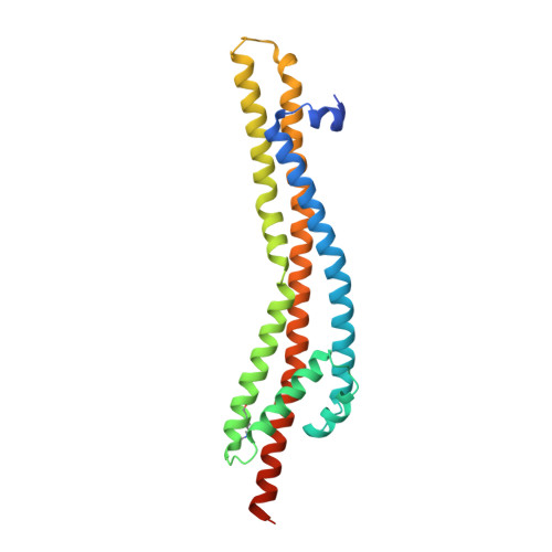
    In Gram-negative bacteria, outer membrane transporters import nutrients by coupling to an inner membrane protein complex called the Ton complex. The Ton complex consists of TonB, ExbB, and ExbD, and uses the proton motive force at the inner membrane to transduce energy to the outer membrane via TonB. Here, we structurally characterize the Ton complex from Escherichia coli using X-ray crystallography, electron microscopy, double electron-electron resonance (DEER) spectroscopy, and crosslinking. Our results reveal a stoichiometry consisting of a pentamer of ExbB, a dimer of ExbD, and at least one TonB. Electrophysiology studies show that the Ton subcomplex forms pH-sensitive cation-selective channels and provide insight into the mechanism by which it may harness the proton motive force to produce energy.


  • Organizational Affiliation

    Laboratoire d'Ingénierie des Systèmes Macromoléculaires, UMR7255 CNRS/Aix-Marseille Université, Institut de Microbiologie de la Méditerranée, 13402 Marseille Cedex 20, France.


Macromolecules
Find similar proteins by:  (by identity cutoff)  |  3D Structure
Entity ID: 1
MoleculeChains Sequence LengthOrganismDetailsImage
Biopolymer transport protein ExbB244Escherichia coli DH1Mutation(s): 0 
Gene Names: exbB
Membrane Entity: Yes 
UniProt
Find proteins for P0ABU7 (Escherichia coli (strain K12))
Explore P0ABU7 
Go to UniProtKB:  P0ABU7
Entity Groups  
Sequence Clusters30% Identity50% Identity70% Identity90% Identity95% Identity100% Identity
UniProt GroupP0ABU7
Sequence Annotations
Expand
  • Reference Sequence
Small Molecules
Modified Residues  1 Unique
IDChains TypeFormula2D DiagramParent
MLY
Query on MLY
A [auth B]
B [auth C]
C [auth D]
D [auth E]
E [auth F]
A [auth B],
B [auth C],
C [auth D],
D [auth E],
E [auth F],
F [auth G],
G [auth H],
H [auth I],
I [auth J],
J [auth A]
L-PEPTIDE LINKINGC8 H18 N2 O2LYS
Experimental Data & Validation

Experimental Data

  • Method: X-RAY DIFFRACTION
  • Resolution: 2.60 Å
  • R-Value Free: 0.258 
  • R-Value Work: 0.211 
  • R-Value Observed: 0.212 
  • Space Group: P 1 21 1
Unit Cell:
Length ( Å )Angle ( ˚ )
a = 137.166α = 90
b = 104.779β = 91.77
c = 149.364γ = 90
Software Package:
Software NamePurpose
PHENIXrefinement
DENZOdata reduction
SCALEPACKdata scaling
PHASERphasing

Structure Validation

View Full Validation Report


Entry History & Funding Information

Deposition Data


Funding OrganizationLocationGrant Number
National Institutes of Health/National Institute Of Allergy and Infectious Diseases (NIH/NIAID)United States1K22AI113078-01
National Institutes of Health/National Institute of Diabetes and Digestive and Kidney Disease (NIH/NIDDK)United StatesZIA DK036139-10
French National Research AgencyFranceANR-14-CE09-0023
Projets internationaux de cooperation scientifiqueFrancePICS05853

Revision History  (Full details and data files)

  • Version 1.0: 2016-09-28
    Type: Initial release
  • Version 1.1: 2016-10-05
    Changes: Database references
  • Version 1.2: 2016-10-19
    Changes: Database references
  • Version 1.3: 2017-09-27
    Changes: Author supporting evidence, Database references, Derived calculations
  • Version 1.4: 2017-10-04
    Changes: Author supporting evidence
  • Version 1.5: 2019-12-11
    Changes: Author supporting evidence