


Crystal Structure of human formylglycine generating enzyme in complex with N-acetyl-cysteine methylester
Kowal, J., Schlotawa, L., Rudolph, M.G., Niemann, H.To be published.
Experimental Data Snapshot
Starting Model: other
View more details
Entity ID: 1 | |||||
|---|---|---|---|---|---|
| Molecule | Chains | Sequence Length | Organism | Details | Image |
| Formylglycine-generating enzyme | 323 | Homo sapiens | Mutation(s): 0 Gene Names: SUMF1, PSEC0152, UNQ3037/PRO9852 EC: 1.8.3.7 | 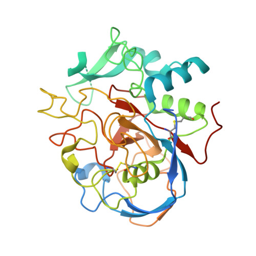 | |
UniProt & NIH Common Fund Data Resources | |||||
PHAROS: Q8NBK3 GTEx: ENSG00000144455 | |||||
Entity Groups | |||||
| Sequence Clusters | 30% Identity50% Identity70% Identity90% Identity95% Identity100% Identity | ||||
| UniProt Group | Q8NBK3 | ||||
Glycosylation | |||||
| Glycosylation Sites: 1 | Go to GlyGen: Q8NBK3-1 | ||||
Sequence AnnotationsExpand | |||||
Reference Sequence | |||||
| Ligands 4 Unique | |||||
|---|---|---|---|---|---|
| ID | Chains | Name / Formula / InChI Key | 2D Diagram | 3D Interactions | |
| NAG Download:Ideal Coordinates CCD File | C [auth A] | 2-acetamido-2-deoxy-beta-D-glucopyranose C8 H15 N O6 OVRNDRQMDRJTHS-FMDGEEDCSA-N |  | ||
| UG6 (Subject of Investigation/LOI) Download:Ideal Coordinates CCD File | F [auth A] | methyl N-acetyl-L-cysteinate C6 H11 N O3 S QTKAQJWFVXPIFV-YFKPBYRVSA-N |  | ||
| CU1 Download:Ideal Coordinates CCD File | B [auth A] | COPPER (I) ION Cu VMQMZMRVKUZKQL-UHFFFAOYSA-N |  | ||
| CA Download:Ideal Coordinates CCD File | D [auth A], E [auth A] | CALCIUM ION Ca BHPQYMZQTOCNFJ-UHFFFAOYSA-N |  | ||
| Length ( Å ) | Angle ( ˚ ) |
|---|---|
| a = 43.475 | α = 90 |
| b = 61.731 | β = 90 |
| c = 109.719 | γ = 90 |
| Software Name | Purpose |
|---|---|
| XSCALE | data scaling |
| PHENIX | refinement |
| PDB_EXTRACT | data extraction |
| XDS | data reduction |
| PHASER | phasing |
| Funding Organization | Location | Grant Number |
|---|---|---|
| University of Bielefeld | Germany | -- |


