


PanDDA analysis group deposition
Imprachim, N., Yosaatmadja, Y., von-Delft, F., Bountra, C., Gileadi, O., Newman, J.A.To be published.
Experimental Data Snapshot
Starting Model: experimental
View more details
Entity ID: 1 | |||||
|---|---|---|---|---|---|
| Molecule | Chains | Sequence Length | Organism | Details | Image |
| Proofreading exoribonuclease nsp14 | A [auth D] | 523 | Severe acute respiratory syndrome coronavirus 2 | Mutation(s): 0 Gene Names: rep, 1a-1b EC: 3.1.13 | 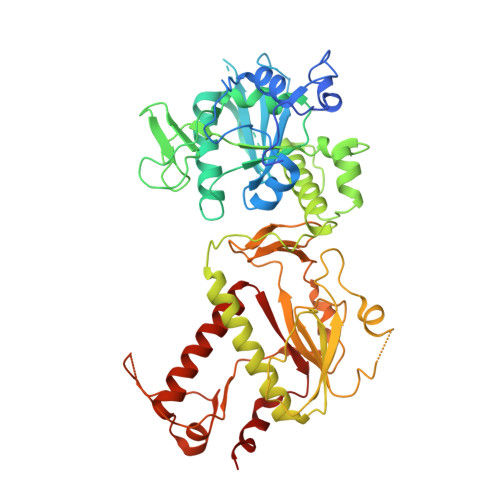 |
UniProt | |||||
Entity Groups | |||||
| Sequence Clusters | 30% Identity50% Identity70% Identity90% Identity95% Identity100% Identity | ||||
| UniProt Group | P0DTD1 | ||||
Sequence AnnotationsExpand | |||||
Reference Sequence | |||||
| Ligands 3 Unique | |||||
|---|---|---|---|---|---|
| ID | Chains | Name / Formula / InChI Key | 2D Diagram | 3D Interactions | |
| 60P (Subject of Investigation/LOI) Download:Ideal Coordinates CCD File | G [auth D], H [auth D] | 3-methylthiophene-2-carboxylic acid C6 H6 O2 S IFLKEBSJTZGCJG-UHFFFAOYSA-N |  | ||
| PO4 Download:Ideal Coordinates CCD File | E [auth D], F [auth D] | PHOSPHATE ION O4 P NBIIXXVUZAFLBC-UHFFFAOYSA-K |  | ||
| ZN Download:Ideal Coordinates CCD File | B [auth D], C [auth D], D | ZINC ION Zn PTFCDOFLOPIGGS-UHFFFAOYSA-N |  | ||
| Length ( Å ) | Angle ( ˚ ) |
|---|---|
| a = 68.125 | α = 90 |
| b = 67.928 | β = 90 |
| c = 138.368 | γ = 90 |
| Software Name | Purpose |
|---|---|
| BUSTER | refinement |
| Aimless | data scaling |
| PDB_EXTRACT | data extraction |
| XDS | data reduction |
| REFMAC | phasing |
| Funding Organization | Location | Grant Number |
|---|---|---|
| Not funded | United Kingdom | -- |


