


Crystal structure of Endothiapepsin
Huschmann, F.U., Weiss, M.S., Mueller, U., Haustedt, L.O., Klebe, G.To be published.
Experimental Data Snapshot
Entity ID: 1 | |||||
|---|---|---|---|---|---|
| Molecule | Chains | Sequence Length | Organism | Details | Image |
| Endothiapepsin | 330 | Cryphonectria parasitica | Mutation(s): 0 EC: 3.4.23.22 | 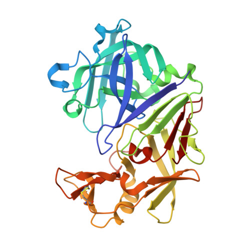 | |
UniProt | |||||
Entity Groups | |||||
| Sequence Clusters | 30% Identity50% Identity70% Identity90% Identity95% Identity100% Identity | ||||
| UniProt Group | P11838 | ||||
Sequence AnnotationsExpand | |||||
Reference Sequence | |||||
| Ligands 5 Unique | |||||
|---|---|---|---|---|---|
| ID | Chains | Name / Formula / InChI Key | 2D Diagram | 3D Interactions | |
| D9D Download:Ideal Coordinates CCD File | F [auth A], G [auth A] | (1S,2S,3S,4R,5R)-2-({[5-(hydroxymethyl)furan-2-yl]methyl}amino)-4-(morpholin-4-yl)-6,8-dioxabicyclo[3.2.1]octan-3-ol C16 H24 N2 O6 QPKSUNGYGHKTGL-DGXTUMSLSA-N |  | ||
| GOL Download:Ideal Coordinates CCD File | B [auth A] | GLYCEROL C3 H8 O3 PEDCQBHIVMGVHV-UHFFFAOYSA-N |  | ||
| DMS Download:Ideal Coordinates CCD File | E [auth A] | DIMETHYL SULFOXIDE C2 H6 O S IAZDPXIOMUYVGZ-UHFFFAOYSA-N |  | ||
| EDO Download:Ideal Coordinates CCD File | C [auth A], D [auth A] | 1,2-ETHANEDIOL C2 H6 O2 LYCAIKOWRPUZTN-UHFFFAOYSA-N |  | ||
| ACT Download:Ideal Coordinates CCD File | H [auth A] | ACETATE ION C2 H3 O2 QTBSBXVTEAMEQO-UHFFFAOYSA-M |  | ||
| Length ( Å ) | Angle ( ˚ ) |
|---|---|
| a = 45.479 | α = 90 |
| b = 73.724 | β = 110.23 |
| c = 53.34 | γ = 90 |
| Software Name | Purpose |
|---|---|
| PHENIX | refinement |
| XSCALE | data scaling |
| PDB_EXTRACT | data extraction |


