5OXE

Structure of major capsid protein VP1 of Aeropyrum pernix bacilliform virus 1 APBV1


Experimental Data Snapshot

  • Method: ELECTRON MICROSCOPY
  • Resolution: 3.70 Å
  • Aggregation State: HELICAL ARRAY 
  • Reconstruction Method: SINGLE PARTICLE 

wwPDB Validation   3D Report Full Report


This is version 1.3 of the entry. See complete history


Literature

Unique architecture of thermophilic archaeal virus APBV1 and its genome packaging.

Ptchelkine, D.Gillum, A.Mochizuki, T.Lucas-Staat, S.Liu, Y.Krupovic, M.Phillips, S.E.V.Prangishvili, D.Huiskonen, J.T.

(2017) Nat Commun 8: 1436-1436

  • DOI: https://doi.org/10.1038/s41467-017-01668-0
  • Primary Citation of Related Structures:  
    5OXE

  • PubMed Abstract: 

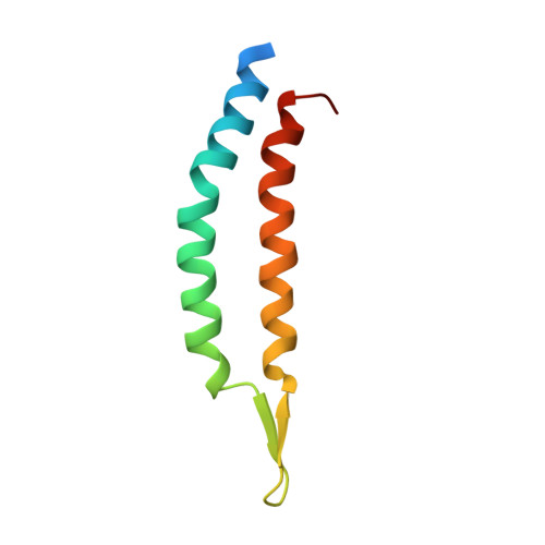
    Archaeal viruses have evolved to infect hosts often thriving in extreme conditions such as high temperatures. However, there is a paucity of information on archaeal virion structures, genome packaging, and determinants of temperature resistance. The rod-shaped virus APBV1 (Aeropyrum pernix bacilliform virus 1) is among the most thermostable viruses known; it infects a hyperthermophile Aeropyrum pernix, which grows optimally at 90 °C. Here we report the structure of APBV1, determined by cryo-electron microscopy at near-atomic resolution. Tight packing of the major virion glycoprotein (VP1) is ensured by extended hydrophobic interfaces, and likely contributes to the extreme thermostability of the helical capsid. The double-stranded DNA is tightly packed in the capsid as a left-handed superhelix and held in place by the interactions with positively charged residues of VP1. The assembly is closed by specific capping structures at either end, which we propose to play a role in DNA packing and delivery.


  • Organizational Affiliation

    Weatherall Institute of Molecular Medicine, University of Oxford, Headley Way, Oxford, OX3 9DS, UK.


Macromolecules
Find similar proteins by:  (by identity cutoff)  |  3D Structure
Entity ID: 1
MoleculeChains Sequence LengthOrganismDetailsImage
Major virion protein81Aeropyrum pernix bacilliform virus 1 Japan/Tanaka/2005Mutation(s): 0 
UniProt
Find proteins for D4QF72 (Aeropyrum pernix bacilliform virus 1 (isolate -/Japan/Tanaka/2005))
Explore D4QF72 
Go to UniProtKB:  D4QF72
Entity Groups  
Sequence Clusters30% Identity50% Identity70% Identity90% Identity95% Identity100% Identity
UniProt GroupD4QF72
Sequence Annotations
Expand
  • Reference Sequence
Experimental Data & Validation

Experimental Data

  • Method: ELECTRON MICROSCOPY
  • Resolution: 3.70 Å
  • Aggregation State: HELICAL ARRAY 
  • Reconstruction Method: SINGLE PARTICLE 
EM Software:
TaskSoftware PackageVersion
RECONSTRUCTIONSPRING
MODEL REFINEMENTREFMAC
MODEL REFINEMENTPHENIX

Structure Validation

View Full Validation Report


Entry History & Funding Information

Deposition Data


Funding OrganizationLocationGrant Number
Wellcome TrustUnited Kingdom203141/Z/16/Z
Wellcome TrustUnited Kingdom060208/Z/00/Z
Wellcome TrustUnited Kingdom093305/Z/10/Z
European Research CouncilUnited Kingdom649053

Revision History  (Full details and data files)

  • Version 1.0: 2017-11-22
    Type: Initial release
  • Version 1.1: 2018-10-17
    Changes: Data collection, Refinement description
  • Version 1.2: 2019-11-06
    Changes: Data collection
  • Version 1.3: 2019-12-11
    Changes: Other