Crystal Structure of the Japanese Encephalitis Virus Capsid Protein.
Poonsiri, T., Wright, G.S.A., Solomon, T., Antonyuk, S.V.(2019) Viruses 11
- PubMed: 31284608 Search on PubMedSearch on PubMed Central
- DOI: https://doi.org/10.3390/v11070623
- Primary Citation Related Structures:
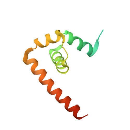
5OW2 - PubMed Abstract:
Japanese encephalitis (JE) is inflammation and swelling of the brain caused by the JE virus (JEV), a mosquito-borne member of the Flavivirus family. There are around 68,000 JE cases worldwide each year, many of which result in permanent brain damage and death. There is no specific treatment for JE. Here we present the crystal structure of the JEV capsid protein, a potential drug target, at 1.98 Å, and compare it to other flavivirus capsid proteins. The JEV capsid has a helical secondary structure (α helixes 1-4) and a similar protein fold to the dengue virus (DENV), the West Nile virus (WNV), and the Zika virus (ZIKV) capsid proteins. It forms a homodimer by antiparallel pairing with another subunit (') through α-helix 1-1', 2-2', and 4-4' interactions. This dimeric form is believed to be the building block of the nucleocapsid. The flexibility of the N-terminal α helix-1 allows the formation of closed and open conformations with possible functional importance. The basic C-terminal pairing of α4-4' forms a coiled-coil-like structure, indicating possible nucleic acid binding functionality. However, a comparison with other nucleic acid interacting domains indicates that homodimerization would preclude binding. This is the first JEV capsid protein to be described and is an addition to the structural biology of the Flavivirus.
- Molecular Biophysics Group, Institute of Integrative Biology, Faculty of Health and Life Sciences, University of Liverpool, L69 7ZB Liverpool, UK.
Organizational Affiliation: