5OSA | pdb_00005osa

GLIC-GABAAR alpha1 chimera crystallized at pH4.6


Experimental Data Snapshot

  • Method: X-RAY DIFFRACTION
  • Resolution: 2.75 Å
  • R-Value Free: 
    0.229 (Depositor), 0.242 (DCC) 
  • R-Value Work: 
    0.203 (Depositor), 0.219 (DCC) 
  • R-Value Observed: 
    0.205 (Depositor) 

Starting Model: experimental
View more details

wwPDB Validation 3D Report Full Report

Validation slider image for 5OSA

Ligand Structure Quality Assessment 


This is version 1.4 of the entry. See complete history

Literature

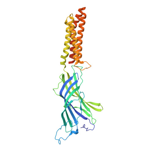
Crystal structures of a GABAA-receptor chimera reveal new endogenous neurosteroid-binding sites.

Laverty, D.Thomas, P.Field, M.Andersen, O.J.Gold, M.G.Biggin, P.C.Gielen, M.Smart, T.G.

(2017) Nat Struct Mol Biol 24: 977-985

  • DOI: https://doi.org/10.1038/nsmb.3477
  • Primary Citation Related Structures: 
    5OSA, 5OSB, 5OSC

  • PubMed Abstract: 

    γ-Aminobutyric acid receptors (GABA A Rs) are vital for controlling excitability in the brain. This is emphasized by the numerous neuropsychiatric disorders that result from receptor dysfunction. A critical component of most native GABA A Rs is the α subunit. Its transmembrane domain is the target for many modulators, including endogenous brain neurosteroids that impact anxiety, stress and depression, and for therapeutic drugs, such as general anesthetics. Understanding the basis for the modulation of GABA A R function requires high-resolution structures. Here we present the first atomic structures of a GABA A R chimera at 2.8-Å resolution, including those bound with potentiating and inhibitory neurosteroids. These structures define new allosteric binding sites for these modulators that are associated with the α-subunit transmembrane domain. Our findings will enable the exploitation of neurosteroids for therapeutic drug design to regulate GABA A Rs in neurological disorders.


  • Organizational Affiliation
    • Department of Neuroscience, Physiology & Pharmacology, University College London, London, UK.

Macromolecule Content 

  • Total Structure Weight: 197.74 kDa 
  • Atom Count: 12,802 
  • Modeled Residue Count: 1,565 
  • Deposited Residue Count: 1,680 
  • Unique protein chains: 1

Macromolecules

Find similar proteins by:|  3D Structure
Entity ID: 1
MoleculeChains  Sequence LengthOrganismDetailsImage
Proton-gated ion channel,Gamma-aminobutyric acid receptor subunit alpha-1,Gamma-aminobutyric acid receptor subunit alpha-1
A, B, C, D, E
336Gloeobacter violaceus PCC 7421Mus musculusMutation(s): 0 
Gene Names: glvIglr4197Gabra1Gabra-1
Membrane Entity: Yes 
UniProt
Find proteins for P62812 (Mus musculus)
Explore P62812 
Go to UniProtKB:  P62812
Find proteins for Q7NDN8 (Gloeobacter violaceus (strain ATCC 29082 / PCC 7421))
Explore Q7NDN8 
Go to UniProtKB:  Q7NDN8
Entity Groups
Sequence Clusters30% Identity50% Identity70% Identity90% Identity95% Identity100% Identity
UniProt GroupsQ7NDN8P62812
Sequence Annotations
Expand
Reference Sequence

Small Molecules

Ligands 4 Unique
IDChains Name / Formula / InChI Key2D Diagram3D Interactions
Y01

Query on Y01


Download:Ideal Coordinates CCD File
BA [auth D],
GA [auth E],
L [auth A],
Q [auth B],
V [auth C]
CHOLESTEROL HEMISUCCINATE
C31 H50 O4
WLNARFZDISHUGS-MIXBDBMTSA-N
D12

Query on D12


Download:Ideal Coordinates CCD File
AA [auth D]
EA [auth E]
FA [auth E]
K [auth A]
O [auth B]
AA [auth D],
EA [auth E],
FA [auth E],
K [auth A],
O [auth B],
P [auth B],
U [auth C],
Z [auth D]
DODECANE
C12 H26
SNRUBQQJIBEYMU-UHFFFAOYSA-N
ACT

Query on ACT


Download:Ideal Coordinates CCD File
CA [auth E]
F [auth A]
G [auth A]
M [auth B]
R [auth C]
CA [auth E],
F [auth A],
G [auth A],
M [auth B],
R [auth C],
S [auth C],
W [auth D],
X [auth D]
ACETATE ION
C2 H3 O2
QTBSBXVTEAMEQO-UHFFFAOYSA-M
CL

Query on CL


Download:Ideal Coordinates CCD File
DA [auth E]
H [auth A]
I [auth A]
J [auth A]
N [auth B]
DA [auth E],
H [auth A],
I [auth A],
J [auth A],
N [auth B],
T [auth C],
Y [auth D]
CHLORIDE ION
Cl
VEXZGXHMUGYJMC-UHFFFAOYSA-M

Experimental Data & Validation

Experimental Data

  • Method: X-RAY DIFFRACTION
  • Resolution: 2.75 Å
  • R-Value Free:  0.229 (Depositor), 0.242 (DCC) 
  • R-Value Work:  0.203 (Depositor), 0.219 (DCC) 
  • R-Value Observed: 0.205 (Depositor) 
Space Group: C 1 2 1
Unit Cell:
Length ( Å )Angle ( ˚ )
a = 185.017α = 90
b = 133.947β = 103.535
c = 162.697γ = 90
Software Package:
Software NamePurpose
BUSTERrefinement
PHENIXrefinement
xia2data processing
PHASERphasing
BUCCANEERmodel building
xia2data reduction
Aimlessdata scaling

Structure Validation

View Full Validation Report


Ligand Structure Quality Assessment 


Entry History 

Deposition Data

Revision History  (Full details and data files)

  • Version 1.0: 2017-10-11
    Type: Initial release
  • Version 1.1: 2017-11-15
    Changes: Database references
  • Version 1.2: 2018-01-31
    Changes: Database references
  • Version 1.3: 2019-10-30
    Changes: Data collection, Database references, Source and taxonomy, Structure summary
  • Version 1.4: 2024-01-17
    Changes: Data collection, Database references, Refinement description