5ORQ

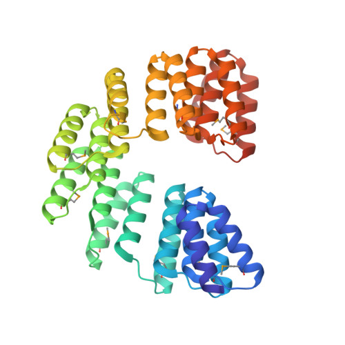
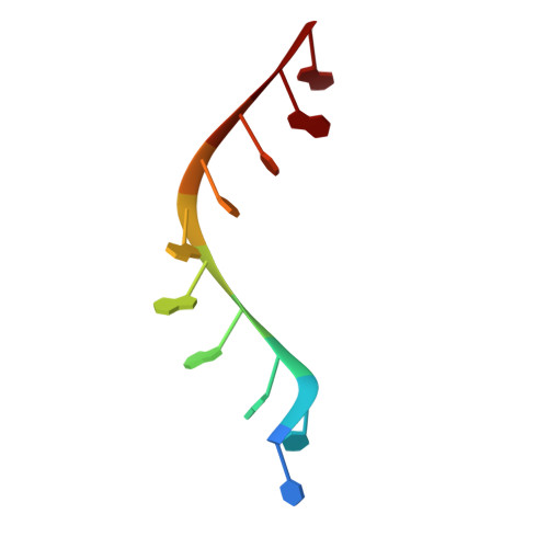
Crystal structure of designed cPPR-Telo1 in complex with ssDNA


Experimental Data Snapshot

  • Method: X-RAY DIFFRACTION
  • Resolution: 1.95 Å
  • R-Value Free: 0.228 
  • R-Value Work: 0.189 
  • R-Value Observed: 0.191 

wwPDB Validation   3D Report Full Report


This is version 1.0 of the entry. See complete history


Literature

Modular ssDNA binding and inhibition of telomerase activity by designer PPR proteins.

Spahr, H.Chia, T.Lingford, J.P.Siira, S.J.Cohen, S.B.Filipovska, A.Rackham, O.

(2018) Nat Commun 9: 2212-2212

  • DOI: https://doi.org/10.1038/s41467-018-04388-1
  • Primary Citation of Related Structures:  
    5ORM, 5ORQ

  • PubMed Abstract: 

    DNA is typically found as a double helix, however it must be separated into single strands during all phases of DNA metabolism; including transcription, replication, recombination and repair. Although recent breakthroughs have enabled the design of modular RNA- and double-stranded DNA-binding proteins, there are currently no tools available to manipulate single-stranded DNA (ssDNA). Here we show that artificial pentatricopeptide repeat (PPR) proteins can be programmed for sequence-specific ssDNA binding. Interactions occur using the same code and specificity as for RNA binding. We solve the structures of DNA-bound and apo proteins revealing the basis for ssDNA binding and how hydrogen bond rearrangements enable the PPR structure to envelope its ssDNA target. Finally, we show that engineered PPRs can be designed to bind telomeric ssDNA and can block telomerase activity. The modular mode of ssDNA binding by PPR proteins provides tools to target ssDNA and to understand its importance in cells.


  • Organizational Affiliation

    Department of Mitochondrial Biology, Max Planck Institute for Biology of Ageing, D-50931, Cologne, Germany.


Macromolecules

Find similar proteins by:  (by identity cutoff)  |  3D Structure
Entity ID: 1
MoleculeChains Sequence LengthOrganismDetailsImage
cPPR-Telo1364synthetic constructMutation(s): 0 
Entity Groups  
Sequence Clusters30% Identity50% Identity70% Identity90% Identity95% Identity100% Identity
Sequence Annotations
Expand
  • Reference Sequence

Find similar nucleic acids by:  Sequence   |   3D Structure  

Entity ID: 2
MoleculeChains LengthOrganismImage
DNA (5'-D(P*TP*TP*AP*GP*GP*GP*TP*TP*AP*G)-3')10Homo sapiens
Sequence Annotations
Expand
  • Reference Sequence
Small Molecules
Modified Residues  1 Unique
IDChains TypeFormula2D DiagramParent
MSE
Query on MSE
A
L-PEPTIDE LINKINGC5 H11 N O2 SeMET
Experimental Data & Validation

Experimental Data

  • Method: X-RAY DIFFRACTION
  • Resolution: 1.95 Å
  • R-Value Free: 0.228 
  • R-Value Work: 0.189 
  • R-Value Observed: 0.191 
  • Space Group: P 41 21 2
Unit Cell:
Length ( Å )Angle ( ˚ )
a = 114.831α = 90
b = 114.831β = 90
c = 83.545γ = 90
Software Package:
Software NamePurpose
Aimlessdata scaling
BUSTERrefinement
PDB_EXTRACTdata extraction
XDSdata reduction
autoSHARPphasing

Structure Validation

View Full Validation Report


Entry History & Funding Information

Deposition Data


Funding OrganizationLocationGrant Number
Australian Research CouncilAustraliaFT0991008, FT0991113, DP140104111
National Health and Medical Research CouncilAustraliaAPP1058442, APP1045677
Cancer Council Western AustraliaAustralia--

Revision History  (Full details and data files)

  • Version 1.0: 2018-06-20
    Type: Initial release