5OQ2

Se-SAD structure of the functional region of Cwp19 from Clostridium difficile


Experimental Data Snapshot

  • Method: X-RAY DIFFRACTION
  • Resolution: 2.30 Å
  • R-Value Free: 0.254 
  • R-Value Work: 0.193 
  • R-Value Observed: 0.196 

wwPDB Validation   3D Report Full Report


This is version 1.2 of the entry. See complete history


Literature

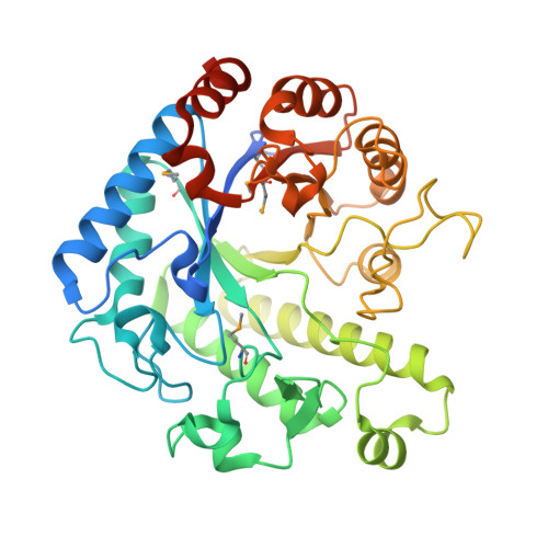
The molecular structure of the glycoside hydrolase domain of Cwp19 from Clostridium difficile.

Bradshaw, W.J.Kirby, J.M.Roberts, A.K.Shone, C.C.Acharya, K.R.

(2017) FEBS J 284: 4343-4357

  • DOI: https://doi.org/10.1111/febs.14310
  • Primary Citation of Related Structures:  
    5OQ2, 5OQ3

  • PubMed Abstract: 

    Clostridium difficile is a burden to healthcare systems around the world, causing tens of thousands of deaths annually. The S-layer of the bacterium, a layer of protein found of the surface of cells, has received a significant amount of attention over the past two decades as a potential target to combat the growing threat presented by C. difficile infections. The S-layer contains a wide range of proteins, each of which possesses three cell wall-binding domains, while many also possess a "functional" region. Here, we present the high resolution structure of the functional region of one such protein, Cwp19 along with preliminary functional characterisation of the predicted glycoside hydrolase. Cwp19 has a TIM barrel fold and appears to possess a high degree of substrate selectivity. The protein also exhibits peptidoglycan hydrolase activity, an order of magnitude slower than that of lysozyme and is the first member of glycoside hydrolase-like family 10 to be characterised. This research goes some way to understanding the role of Cwp19 in the S-layer of C. difficile.


  • Organizational Affiliation

    Department of Biology and Biochemistry, University of Bath, UK.


Macromolecules
Find similar proteins by:  (by identity cutoff)  |  3D Structure
Entity ID: 1
MoleculeChains Sequence LengthOrganismDetailsImage
Cwp19
A, B
396Clostridioides difficileMutation(s): 0 
Gene Names: lytC_21cwp19lytC_5SAMEA3374989_00994SAMEA3375004_02322
EC: 3.5.1.28
UniProt
Find proteins for L7PGA3 (Clostridioides difficile)
Explore L7PGA3 
Go to UniProtKB:  L7PGA3
Entity Groups  
Sequence Clusters30% Identity50% Identity70% Identity90% Identity95% Identity100% Identity
UniProt GroupL7PGA3
Sequence Annotations
Expand
  • Reference Sequence
Small Molecules
Modified Residues  1 Unique
IDChains TypeFormula2D DiagramParent
MSE
Query on MSE
A, B
L-PEPTIDE LINKINGC5 H11 N O2 SeMET
Experimental Data & Validation

Experimental Data

  • Method: X-RAY DIFFRACTION
  • Resolution: 2.30 Å
  • R-Value Free: 0.254 
  • R-Value Work: 0.193 
  • R-Value Observed: 0.196 
  • Space Group: P 1 21 1
Unit Cell:
Length ( Å )Angle ( ˚ )
a = 55.28α = 90
b = 60.41β = 94.21
c = 105.05γ = 90
Software Package:
Software NamePurpose
REFMACrefinement
XDSdata reduction
XSCALEdata scaling
CRANK2phasing

Structure Validation

View Full Validation Report


Entry History & Funding Information

Deposition Data


Funding OrganizationLocationGrant Number
Medical Research Council (United Kingdom)MR/K027123/1

Revision History  (Full details and data files)

  • Version 1.0: 2017-11-01
    Type: Initial release
  • Version 1.1: 2017-11-08
    Changes: Database references
  • Version 1.2: 2017-12-27
    Changes: Database references