5OJ9 | pdb_00005oj9

Structure of Mb NMH


Experimental Data Snapshot

  • Method: X-RAY DIFFRACTION
  • Resolution: 1.48 Å
  • R-Value Free: 
    0.232 (Depositor), 0.231 (DCC) 
  • R-Value Work: 
    0.200 (Depositor), 0.200 (DCC) 
  • R-Value Observed: 
    0.201 (Depositor) 

Starting Model: experimental
View more details

wwPDB Validation 3D Report Full Report

Validation slider image for 5OJ9

Ligand Structure Quality Assessment 


This is version 1.2 of the entry. See complete history

Literature

A Noncanonical Proximal Heme Ligand Affords an Efficient Peroxidase in a Globin Fold.

Pott, M.Hayashi, T.Mori, T.Mittl, P.R.E.Green, A.P.Hilvert, D.

(2018) J Am Chem Soc 140: 1535-1543

  • DOI: https://doi.org/10.1021/jacs.7b12621
  • Primary Citation Related Structures: 
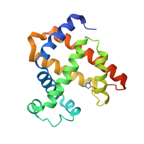
    5OJ9, 5OJA, 5OJB, 5OJC

  • PubMed Abstract: 

    Expanding the range of genetically encoded metal coordination environments accessible within tunable protein scaffolds presents excellent opportunities for the creation of metalloenzymes with augmented properties and novel activities. Here, we demonstrate that installation of a noncanonical N δ -methyl histidine (NMH) as the proximal heme ligand in the oxygen binding protein myoglobin (Mb) leads to substantial increases in heme redox potential and promiscuous peroxidase activity. Structural characterization of this catalytically modified myoglobin variant (Mb NMH) revealed significant changes in the proximal pocket, including alterations to hydrogen-bonding interactions involving the prosthetic porphyrin cofactor. Further optimization of Mb NMH via a combination of rational modification and several rounds of laboratory evolution afforded efficient peroxidase biocatalysts within a globin fold, with activities comparable to those displayed by nature's peroxidases.


  • Organizational Affiliation
    • Laboratory of Organic Chemistry, ETH Zurich , Zurich 8093, Switzerland.

Macromolecule Content 

  • Total Structure Weight: 19.03 kDa 
  • Atom Count: 1,401 
  • Modeled Residue Count: 154 
  • Deposited Residue Count: 163 
  • Unique protein chains: 1

Macromolecules

Find similar proteins by:|  3D Structure
Entity ID: 1
MoleculeChains  Sequence LengthOrganismDetailsImage
Myoglobin163Physeter macrocephalusMutation(s): 0 
Gene Names: MB
EC: 1.11.1 (UniProt), 1.7 (UniProt)
UniProt
Find proteins for P02185 (Physeter macrocephalus)
Explore P02185 
Go to UniProtKB:  P02185
Entity Groups
Sequence Clusters30% Identity50% Identity70% Identity90% Identity95% Identity100% Identity
UniProt GroupP02185
Sequence Annotations
Expand
Reference Sequence

Small Molecules

Ligands 1 Unique
IDChains Name / Formula / InChI Key2D Diagram3D Interactions
HEM

Query on HEM


Download:Ideal Coordinates CCD File
B [auth A]PROTOPORPHYRIN IX CONTAINING FE
C34 H32 Fe N4 O4
KABFMIBPWCXCRK-RGGAHWMASA-L
Modified Residues  1 Unique
IDChains TypeFormula2D DiagramParent
MHS
Query on MHS
A
L-PEPTIDE LINKINGC7 H11 N3 O2HIS

Experimental Data & Validation

Experimental Data

  • Method: X-RAY DIFFRACTION
  • Resolution: 1.48 Å
  • R-Value Free:  0.232 (Depositor), 0.231 (DCC) 
  • R-Value Work:  0.200 (Depositor), 0.200 (DCC) 
  • R-Value Observed: 0.201 (Depositor) 
Space Group: P 21 21 21
Unit Cell:
Length ( Å )Angle ( ˚ )
a = 39.712α = 90
b = 47.786β = 90
c = 77.484γ = 90
Software Package:
Software NamePurpose
PHENIXrefinement
XDSdata reduction
XDSdata scaling
PHASERphasing

Structure Validation

View Full Validation Report


Ligand Structure Quality Assessment 


Entry History 

Deposition Data

Revision History  (Full details and data files)

  • Version 1.0: 2018-01-24
    Type: Initial release
  • Version 1.1: 2018-02-07
    Changes: Database references
  • Version 1.2: 2024-01-17
    Changes: Data collection, Database references, Refinement description