5OJ2

Crystal structure of the chicken MDGA1 ectodomain

  • Classification: CELL ADHESION
  • Organism(s): Gallus gallus
  • Expression System: Homo sapiens
  • Mutation(s): Yes 

  • Deposited: 2017-07-20 Released: 2017-08-23 
  • Deposition Author(s): Elegheert, J., Clayton, A.J., Aricescu, A.R.
  • Funding Organization(s): Medical Research Council (United Kingdom), CIHR, National Institutes of Health, European Molecular Biology Organization, Marie-Curie, Cancer Research UK, Wellcome Trust

Experimental Data Snapshot

  • Method: X-RAY DIFFRACTION
  • Resolution: 3.20 Å
  • R-Value Free: 0.251 
  • R-Value Work: 0.214 
  • R-Value Observed: 0.216 

wwPDB Validation   3D Report Full Report


Ligand Structure Quality Assessment 


This is version 2.0 of the entry. See complete history


Literature

Structural Mechanism for Modulation of Synaptic Neuroligin-Neurexin Signaling by MDGA Proteins.

Elegheert, J.Cvetkovska, V.Clayton, A.J.Heroven, C.Vennekens, K.M.Smukowski, S.N.Regan, M.C.Jia, W.Smith, A.C.Furukawa, H.Savas, J.N.de Wit, J.Begbie, J.Craig, A.M.Aricescu, A.R.

(2017) Neuron 95: 896-913.e10

  • DOI: https://doi.org/10.1016/j.neuron.2017.07.040
  • Primary Citation of Related Structures:  
    5OJ2, 5OJ6, 5OJK

  • PubMed Abstract: 

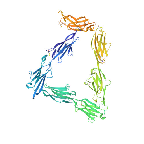
    Neuroligin-neurexin (NL-NRX) complexes are fundamental synaptic organizers in the central nervous system. An accurate spatial and temporal control of NL-NRX signaling is crucial to balance excitatory and inhibitory neurotransmission, and perturbations are linked with neurodevelopmental and psychiatric disorders. MDGA proteins bind NLs and control their function and interaction with NRXs via unknown mechanisms. Here, we report crystal structures of MDGA1, the NL1-MDGA1 complex, and a spliced NL1 isoform. Two large, multi-domain MDGA molecules fold into rigid triangular structures, cradling a dimeric NL to prevent NRX binding. Structural analyses guided the discovery of a broad, splicing-modulated interaction network between MDGA and NL family members and helped rationalize the impact of autism-linked mutations. We demonstrate that expression levels largely determine whether MDGAs act selectively or suppress the synapse organizing function of multiple NLs. These results illustrate a potentially brain-wide regulatory mechanism for NL-NRX signaling modulation.


  • Organizational Affiliation

    Division of Structural Biology, Wellcome Trust Centre for Human Genetics, University of Oxford, Roosevelt Drive, Oxford OX3 7BN, UK. Electronic address: jelegheert@strubi.ox.ac.uk.


Macromolecules
Find similar proteins by:  (by identity cutoff)  |  3D Structure
Entity ID: 1
MoleculeChains Sequence LengthOrganismDetailsImage
MAM domain-containing glycosylphosphatidylinositol anchor protein 1
A, B
910Gallus gallusMutation(s): 1 
Gene Names: MDGA1
UniProt
Find proteins for Q0WYX8 (Gallus gallus)
Explore Q0WYX8 
Go to UniProtKB:  Q0WYX8
Entity Groups  
Sequence Clusters30% Identity50% Identity70% Identity90% Identity95% Identity100% Identity
UniProt GroupQ0WYX8
Sequence Annotations
Expand
  • Reference Sequence
Oligosaccharides

Help

Entity ID: 2
MoleculeChains Length2D Diagram Glycosylation3D Interactions
beta-D-mannopyranose-(1-4)-2-acetamido-2-deoxy-beta-D-glucopyranose-(1-4)-2-acetamido-2-deoxy-beta-D-glucopyranose
C
3N-Glycosylation
Glycosylation Resources
GlyTouCan:  G15407YE
GlyCosmos:  G15407YE
GlyGen:  G15407YE
Entity ID: 3
MoleculeChains Length2D Diagram Glycosylation3D Interactions
alpha-D-mannopyranose-(1-3)-beta-D-mannopyranose-(1-4)-2-acetamido-2-deoxy-beta-D-glucopyranose-(1-4)-2-acetamido-2-deoxy-beta-D-glucopyranose
D
4N-Glycosylation
Glycosylation Resources
GlyTouCan:  G81315DD
GlyCosmos:  G81315DD
GlyGen:  G81315DD
Small Molecules
Ligands 1 Unique
IDChains Name / Formula / InChI Key2D Diagram3D Interactions
NAG
Query on NAG

Download Ideal Coordinates CCD File 
E [auth A]
F [auth A]
G [auth A]
H [auth A]
I [auth A]
E [auth A],
F [auth A],
G [auth A],
H [auth A],
I [auth A],
J [auth A],
K [auth A],
L [auth A],
M [auth B],
N [auth B],
O [auth B],
P [auth B],
Q [auth B],
R [auth B],
S [auth B]
2-acetamido-2-deoxy-beta-D-glucopyranose
C8 H15 N O6
OVRNDRQMDRJTHS-FMDGEEDCSA-N
Modified Residues  1 Unique
IDChains TypeFormula2D DiagramParent
MSE
Query on MSE
A, B
L-PEPTIDE LINKINGC5 H11 N O2 SeMET
Experimental Data & Validation

Experimental Data

  • Method: X-RAY DIFFRACTION
  • Resolution: 3.20 Å
  • R-Value Free: 0.251 
  • R-Value Work: 0.214 
  • R-Value Observed: 0.216 
  • Space Group: P 21 21 21
Unit Cell:
Length ( Å )Angle ( ˚ )
a = 103.18α = 90
b = 109.04β = 90
c = 208.77γ = 90
Software Package:
Software NamePurpose
PHENIXrefinement
xia2data reduction
Aimlessdata scaling
PHENIXphasing

Structure Validation

View Full Validation Report


Ligand Structure Quality Assessment 


Entry History & Funding Information

Deposition Data


Funding OrganizationLocationGrant Number
Medical Research Council (United Kingdom)United KingdomG0700232
Medical Research Council (United Kingdom)United KingdomL009609
Medical Research Council (United Kingdom)United KingdomMC_UP_1201/15
CIHRCanadaFDN-143206
CIHRCanadaNDD-144222
National Institutes of HealthUnited StatesR00 DC013805
European Molecular Biology OrganizationUnited KingdomALTF 1116-2012
Marie-CurieUnited KingdomFP7-328531
Cancer Research UKUnited KingdomC375/A10976
National Institutes of HealthUnited StatesMH085926
National Institutes of HealthUnited StatesGM105730
Wellcome TrustUnited Kingdom090532/Z/09/Z

Revision History  (Full details and data files)

  • Version 1.0: 2017-08-23
    Type: Initial release
  • Version 1.1: 2017-08-30
    Changes: Database references
  • Version 2.0: 2020-07-29
    Type: Remediation
    Reason: Carbohydrate remediation
    Changes: Atomic model, Data collection, Derived calculations, Structure summary