5OIS | pdb_00005ois

InhA (T2A mutant) complexed with 4-((5-Amino-3-methyl-1H-pyrazol-1-yl)methyl)-N-(2-chloro-4-fluorobenzyl)benzamide


Experimental Data Snapshot

  • Method: X-RAY DIFFRACTION
  • Resolution: 2.24 Å
  • R-Value Free: 
    0.189 (Depositor), 0.203 (DCC) 
  • R-Value Work: 
    0.171 (Depositor), 0.180 (DCC) 
  • R-Value Observed: 
    0.172 (Depositor) 

wwPDB Validation 3D Report Full Report

Validation slider image for 5OIS

Ligand Structure Quality Assessment 


This is version 1.2 of the entry. See complete history

Literature

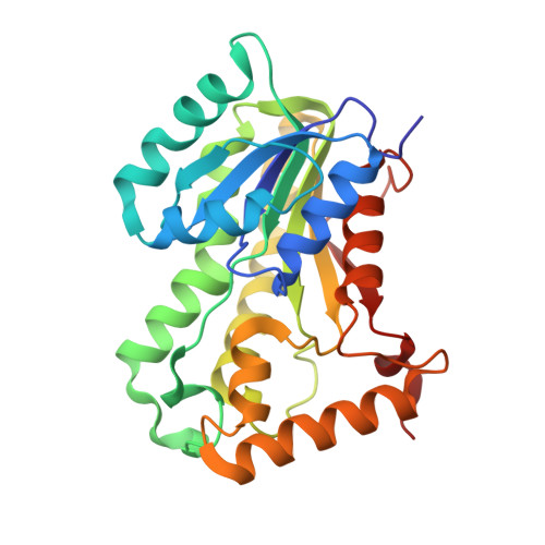
Screening of a Novel Fragment Library with Functional Complexity against Mycobacterium tuberculosis InhA.

Prati, F.Zuccotto, F.Fletcher, D.Convery, M.A.Fernandez-Menendez, R.Bates, R.Encinas, L.Zeng, J.Chung, C.W.De Dios Anton, P.Mendoza-Losana, A.Mackenzie, C.Green, S.R.Huggett, M.Barros, D.Wyatt, P.G.Ray, P.C.

(2018) ChemMedChem 13: 672-677

  • DOI: https://doi.org/10.1002/cmdc.201700774
  • Primary Citation Related Structures: 
    5OIC, 5OIF, 5OIL, 5OIM, 5OIN, 5OIO, 5OIP, 5OIQ, 5OIR, 5OIS, 5OIT

  • PubMed Abstract: 

    Our findings reported herein provide support for the benefits of including functional group complexity (FGC) within fragments when screening against protein targets such as Mycobacterium tuberculosis InhA. We show that InhA fragment actives with FGC maintained their binding pose during elaboration. Furthermore, weak fragment hits with functional group handles also allowed for facile fragment elaboration to afford novel and potent InhA inhibitors with good ligand efficiency metrics for optimization.


  • Organizational Affiliation
    • Drug Discovery Unit, College of Life Sciences, University of Dundee, Dow Street, Dundee, DD1 5EH, Scotland, UK.

Macromolecule Content 

  • Total Structure Weight: 118.24 kDa 
  • Atom Count: 8,720 
  • Modeled Residue Count: 1,072 
  • Deposited Residue Count: 1,076 
  • Unique protein chains: 1

Macromolecules

Find similar proteins by:|  3D Structure
Entity ID: 1
MoleculeChains  Sequence LengthOrganismDetailsImage
Enoyl-[acyl-carrier-protein] reductase [NADH]A [auth B],
B [auth D],
C [auth F],
D [auth H]
269Mycobacterium tuberculosis H37RvMutation(s): 0 
Gene Names: inhARv1484MTCY277.05
EC: 1.3.1.9
UniProt
Find proteins for P9WGR1 (Mycobacterium tuberculosis (strain ATCC 25618 / H37Rv))
Explore P9WGR1 
Go to UniProtKB:  P9WGR1
Entity Groups
Sequence Clusters30% Identity50% Identity70% Identity90% Identity95% Identity100% Identity
UniProt GroupP9WGR1
Sequence Annotations
Expand
Reference Sequence

Small Molecules

Ligands 2 Unique
IDChains Name / Formula / InChI Key2D Diagram3D Interactions
NAD

Query on NAD


Download:Ideal Coordinates CCD File
F [auth B],
H [auth D],
J [auth F],
L [auth H]
NICOTINAMIDE-ADENINE-DINUCLEOTIDE
C21 H27 N7 O14 P2
BAWFJGJZGIEFAR-NNYOXOHSSA-N
9WK
(Subject of Investigation/LOI)

Query on 9WK


Download:Ideal Coordinates CCD File
E [auth B],
G [auth D],
I [auth F],
K [auth H]
4-[(5-azanyl-3-methyl-pyrazol-1-yl)methyl]-~{N}-[(2-chloranyl-4-fluoranyl-phenyl)methyl]benzamide
C19 H18 Cl F N4 O
HOVSFDUYYXXWGJ-UHFFFAOYSA-N
Binding Affinity Annotations 
IDSourceBinding Affinity
9WK BindingDB:  5OIS Kd: 8000 (nM) from 1 assay(s)

Experimental Data & Validation

Experimental Data

  • Method: X-RAY DIFFRACTION
  • Resolution: 2.24 Å
  • R-Value Free:  0.189 (Depositor), 0.203 (DCC) 
  • R-Value Work:  0.171 (Depositor), 0.180 (DCC) 
  • R-Value Observed: 0.172 (Depositor) 
Space Group: P 21 21 21
Unit Cell:
Length ( Å )Angle ( ˚ )
a = 105.54α = 90
b = 103.51β = 90
c = 177.22γ = 90
Software Package:
Software NamePurpose
BUSTERrefinement
XDSdata reduction
Aimlessdata scaling
BUSTERphasing

Structure Validation

View Full Validation Report


Ligand Structure Quality Assessment 


Entry History 

Deposition Data

Revision History  (Full details and data files)

  • Version 1.0: 2018-02-14
    Type: Initial release
  • Version 1.1: 2018-04-18
    Changes: Data collection, Database references
  • Version 1.2: 2024-05-08
    Changes: Data collection, Database references