Insights into the roles of non-catalytic residues in the active site of a GH10 xylanase with activity on cellulose.
Chu, Y., Tu, T., Penttinen, L., Xue, X., Wang, X., Yi, Z., Gong, L., Rouvinen, J., Luo, H., Hakulinen, N., Yao, B., Su, X.(2017) J Biological Chem 292: 19315-19327
- PubMed: 28974575 Search on PubMedSearch on PubMed Central
- DOI: https://doi.org/10.1074/jbc.M117.807768
- Primary Citation Related Structures:
5OFJ, 5OFK, 5OFL - PubMed Abstract:
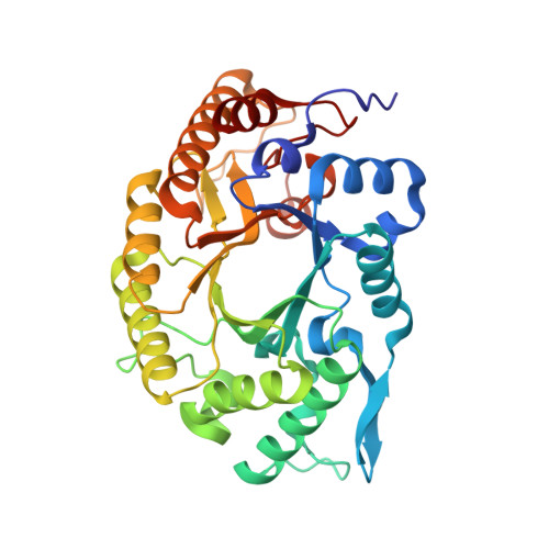
Bifunctional glycoside hydrolases have potential for cost-savings in enzymatic decomposition of plant cell wall polysaccharides for biofuels and bio-based chemicals. The N-terminal GH10 domain of a bifunctional multimodular enzyme Cb Xyn10C/Cel48B from Caldicellulosiruptor bescii is an enzyme able to degrade xylan and cellulose simultaneously. However, the molecular mechanism underlying its substrate promiscuity has not been elucidated. Herein, we discovered that the binding cleft of Cb Xyn10C would have at least six sugar-binding subsites by using isothermal titration calorimetry analysis of the inactive E140Q/E248Q mutant with xylo- and cello-oligosaccharides. This was confirmed by determining the catalytic efficiency of the wild-type enzyme on these oligosaccharides. The free form and complex structures of Cb Xyn10C with xylose- or glucose-configured oligosaccharide ligands were further obtained by crystallographic analysis and molecular modeling and docking. Cb Xyn10C was found to have a typical (β/α) 8 -TIM barrel fold and "salad-bowl" shape of GH10 enzymes. In complex structures with xylo-oligosaccharides, seven sugar-binding subsites were found, and many residues responsible for substrate interactions were identified. Site-directed mutagenesis indicated that 6 and 10 amino acid residues were key residues for xylan and cellulose hydrolysis, respectively. The most important residues are centered on subsites -2 and -1 near the cleavage site, whereas residues playing moderate roles could be located at more distal regions of the binding cleft. Manipulating the residues interacting with substrates in the distal regions directly or indirectly improved the activity of Cb Xyn10C on xylan and cellulose. Most of the key residues for cellulase activity are conserved across GH10 xylanases. Revisiting randomly selected GH10 enzymes revealed unreported cellulase activity, indicating that the dual function may be a more common phenomenon than has been expected.
- From the Key Laboratory for Feed Biotechnology of the Ministry of Agriculture, Feed Research Institute, Chinese Academy of Agricultural Sciences, Beijing 100081, China.
Organizational Affiliation: