NMR relaxation parameters of methyl groups as a tool to map the interfaces of helix-helix interactions in membrane proteins.
Lesovoy, D.M., Mineev, K.S., Bragin, P.E., Bocharova, O.V., Bocharov, E.V., Arseniev, A.S.(2017) J Biomol NMR 69: 165-179
- PubMed: 29063258 Search on PubMed
- DOI: https://doi.org/10.1007/s10858-017-0146-1
- Primary Citation Related Structures:
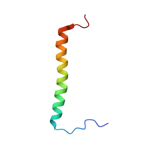
5OB4 - PubMed Abstract:
In the case of soluble proteins, chemical shift mapping is used to identify the intermolecular interfaces when the NOE-based calculations of spatial structure of the molecular assembly are impossible or impracticable. However, the reliability of the membrane protein interface mapping based on chemical shifts or other relevant parameters was never assessed. In the present work, we investigate the predictive power of various NMR parameters that can be used for mapping of helix-helix interfaces in dimeric TM domains. These parameters are studied on a dataset containing three structures of helical dimers obtained for two different proteins in various membrane mimetics. We conclude that the amide chemical shifts have very little predictive value, while the methyl chemical shifts could be used to predict interfaces, though with great care. We suggest an approach based on conversion of the carbon NMR relaxation parameters of methyl groups into parameters of motion, and one of such values, the characteristic time of methyl rotation, appears to be a reliable sensor of interhelix contacts in transmembrane domains. The carbon NMR relaxation parameters of methyl groups can be measured accurately and with high sensitivity and resolution, making the proposed parameter a useful tool for investigation of protein-protein interfaces even in large membrane proteins. An approach to build the models of transmembrane dimers based on perturbations of methyl parameters and TMDOCK software is suggested.
- Shemyakin-Ovchinnikov Institute of Bioorganic Chemistry, Russian Academy of Sciences RAS, Str. Miklukho-Maklaya 16/10, Moscow, Russian Federation, 117997.
Organizational Affiliation: