5O8X

The X-ray Structure of Catenated Lytic Transglycosylase SltB1 from Pseudomonas aeruginosa


Experimental Data Snapshot

  • Method: X-RAY DIFFRACTION
  • Resolution: 2.50 Å
  • R-Value Free: 0.229 
  • R-Value Work: 0.177 
  • R-Value Observed: 0.180 

wwPDB Validation   3D Report Full Report


This is version 1.3 of the entry. See complete history


Literature

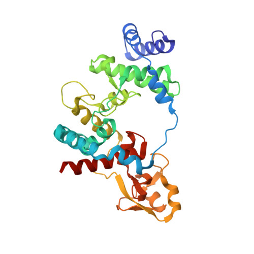
X-ray Structure of Catenated Lytic Transglycosylase SltB1.

Dominguez-Gil, T.Molina, R.Dik, D.A.Spink, E.Mobashery, S.Hermoso, J.A.

(2017) Biochemistry 56: 6317-6320

  • DOI: https://doi.org/10.1021/acs.biochem.7b00932
  • Primary Citation of Related Structures:  
    5O8X

  • PubMed Abstract: 

    Formation of catenanes by proteins is rare, with few known examples. We report herein the X-ray structure of a catenane dimer of lytic transglycosylase SltB1 of Pseudomonas aeruginosa. The enzyme is soluble and exists in the periplasmic space, where it modifies the bacterial cell wall. The catenane dimer exhibits the protein monomers in a noncovalent chain-link arrangement, whereby a stretch of 51 amino acids (to become a loop and three helices) from one monomer threads through the central opening of the structure of the partner monomer. The protein folds after threading in a manner that leaves two helices (α1 and α2) as stoppers to impart stability to the dimer structure. The symmetric embrace by the two SltB1 molecules occludes both active sites entirely, an arrangement that is sustained by six electrostatic interactions between the two monomers. In light of the observation of these structural motifs in all members of Family 3 lytic transglycosylases, catenanes might be present for those enzymes, as well. The dimeric catenane might represent a regulated form of SltB1.


  • Organizational Affiliation

    Department of Crystallography and Structural Biology, Institute of Physical Chemistry "Rocasolano", CSIC , 28006 Madrid, Spain.


Macromolecules
Find similar proteins by:  (by identity cutoff)  |  3D Structure
Entity ID: 1
MoleculeChains Sequence LengthOrganismDetailsImage
Soluble lytic transglycosylase B
A, B
306Pseudomonas aeruginosaMutation(s): 0 
Gene Names: B0B20_29725BZY57_15515BZY58_10660BZY59_02470BZY60_10655
UniProt
Find proteins for Q9HX24 (Pseudomonas aeruginosa (strain ATCC 15692 / DSM 22644 / CIP 104116 / JCM 14847 / LMG 12228 / 1C / PRS 101 / PAO1))
Explore Q9HX24 
Go to UniProtKB:  Q9HX24
Entity Groups  
Sequence Clusters30% Identity50% Identity70% Identity90% Identity95% Identity100% Identity
UniProt GroupQ9HX24
Sequence Annotations
Expand
  • Reference Sequence
Experimental Data & Validation

Experimental Data

  • Method: X-RAY DIFFRACTION
  • Resolution: 2.50 Å
  • R-Value Free: 0.229 
  • R-Value Work: 0.177 
  • R-Value Observed: 0.180 
  • Space Group: C 1 2 1
Unit Cell:
Length ( Å )Angle ( ˚ )
a = 115.064α = 90
b = 116.358β = 118.41
c = 54.865γ = 90
Software Package:
Software NamePurpose
PHENIXrefinement
XDSdata reduction
Aimlessdata scaling
PHASERphasing

Structure Validation

View Full Validation Report


Entry History 

Deposition Data

Revision History  (Full details and data files)

  • Version 1.0: 2017-11-29
    Type: Initial release
  • Version 1.1: 2017-12-13
    Changes: Database references
  • Version 1.2: 2019-10-16
    Changes: Data collection
  • Version 1.3: 2024-05-08
    Changes: Data collection, Database references