5NXL

Formylglycine generating enzyme from T. curvata in complex with Ag(I)


Experimental Data Snapshot

  • Method: X-RAY DIFFRACTION
  • Resolution: 1.66 Å
  • R-Value Free: 0.198 
  • R-Value Work: 0.170 
  • R-Value Observed: 0.172 

wwPDB Validation   3D Report Full Report


This is version 1.2 of the entry. See complete history


Literature

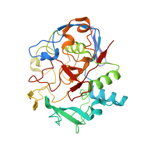
Structural Basis for Copper-Oxygen Mediated C-H Bond Activation by the Formylglycine-Generating Enzyme.

Meury, M.Knop, M.Seebeck, F.P.

(2017) Angew Chem Int Ed Engl 56: 8115-8119

  • DOI: https://doi.org/10.1002/anie.201702901
  • Primary Citation of Related Structures:  
    5NXL, 5NYY

  • PubMed Abstract: 

    The formylglycine-generating enzyme (FGE) is a unique copper protein that catalyzes oxygen-dependent C-H activation. We describe 1.66 Å- and 1.28 Å-resolution crystal structures of FGE from Thermomonospora curvata in complex with either Ag I or Cd II providing definitive evidence for a high-affinity metal-binding site in this enzyme. The structures reveal a bis-cysteine linear coordination of the monovalent metal, and tetrahedral coordination of the bivalent metal. Similar coordination changes may occur in the active enzyme as a result of Cu I/II redox cycling. Complexation of copper atoms by two cysteine residues is common among copper-trafficking proteins, but is unprecedented for redox-active copper enzymes or synthetic copper catalysts.


  • Organizational Affiliation

    Department for Chemistry, University of Basel, St. Johanns-Ring 19, 4056, Basel, Switzerland.


Macromolecules
Find similar proteins by:  (by identity cutoff)  |  3D Structure
Entity ID: 1
MoleculeChains Sequence LengthOrganismDetailsImage
Non-specific serine/threonine protein kinase300Thermomonospora curvata DSM 43183Mutation(s): 0 
Gene Names: Tcur_4811
EC: 2.7.11.1
UniProt
Find proteins for D1A7C3 (Thermomonospora curvata (strain ATCC 19995 / DSM 43183 / JCM 3096 / KCTC 9072 / NBRC 15933 / NCIMB 10081 / Henssen B9))
Explore D1A7C3 
Go to UniProtKB:  D1A7C3
Entity Groups  
Sequence Clusters30% Identity50% Identity70% Identity90% Identity95% Identity100% Identity
UniProt GroupD1A7C3
Sequence Annotations
Expand
  • Reference Sequence
Small Molecules
Ligands 4 Unique
IDChains Name / Formula / InChI Key2D Diagram3D Interactions
MPD
Query on MPD

Download Ideal Coordinates CCD File 
C [auth A](4S)-2-METHYL-2,4-PENTANEDIOL
C6 H14 O2
SVTBMSDMJJWYQN-YFKPBYRVSA-N
AG
Query on AG

Download Ideal Coordinates CCD File 
B [auth A]SILVER ION
Ag
FOIXSVOLVBLSDH-UHFFFAOYSA-N
ACT
Query on ACT

Download Ideal Coordinates CCD File 
D [auth A]
E [auth A]
F [auth A]
G [auth A]
H [auth A]
D [auth A],
E [auth A],
F [auth A],
G [auth A],
H [auth A],
I [auth A],
J [auth A],
K [auth A],
L [auth A],
M [auth A],
N [auth A],
O [auth A]
ACETATE ION
C2 H3 O2
QTBSBXVTEAMEQO-UHFFFAOYSA-M
CA
Query on CA

Download Ideal Coordinates CCD File 
P [auth A],
Q [auth A]
CALCIUM ION
Ca
BHPQYMZQTOCNFJ-UHFFFAOYSA-N
Experimental Data & Validation

Experimental Data

  • Method: X-RAY DIFFRACTION
  • Resolution: 1.66 Å
  • R-Value Free: 0.198 
  • R-Value Work: 0.170 
  • R-Value Observed: 0.172 
  • Space Group: P 21 21 21
Unit Cell:
Length ( Å )Angle ( ˚ )
a = 40.947α = 90
b = 65.572β = 90
c = 99.501γ = 90
Software Package:
Software NamePurpose
PHENIXrefinement
Aimlessdata scaling
PDB_EXTRACTdata extraction
XDSdata reduction
PHASERphasing
XDSdata reduction

Structure Validation

View Full Validation Report


Entry History & Funding Information

Deposition Data


Funding OrganizationLocationGrant Number
European Research Council336559

Revision History  (Full details and data files)

  • Version 1.0: 2017-06-07
    Type: Initial release
  • Version 1.1: 2017-07-05
    Changes: Database references
  • Version 1.2: 2024-01-17
    Changes: Advisory, Author supporting evidence, Data collection, Database references, Derived calculations, Refinement description