5NW7

Crystal structure of candida albicans phosphomannose isomerase in complex with inhibitor


Experimental Data Snapshot

  • Method: X-RAY DIFFRACTION
  • Resolution: 1.85 Å
  • R-Value Free: 0.207 
  • R-Value Work: 0.171 
  • R-Value Observed: 0.173 

wwPDB Validation   3D Report Full Report


Ligand Structure Quality Assessment 


This is version 1.2 of the entry. See complete history


Literature

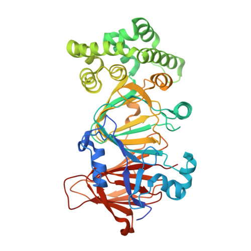
Crystal structure of phosphomannose isomerase from Candida albicans complexed with 5-phospho-d-arabinonhydrazide.

Ahmad, L.Plancqueel, S.Dubosclard, V.Lazar, N.Ghattas, W.Li de la Sierra-Gallay, I.van Tilbeurgh, H.Salmon, L.

(2018) FEBS Lett 592: 1667-1680

  • DOI: https://doi.org/10.1002/1873-3468.13059
  • Primary Citation of Related Structures:  
    5NW7

  • PubMed Abstract: 

    Type I phosphomannose isomerases (PMIs) are zinc-dependent monofunctional metalloenzymes catalysing the reversible isomerization of d-mannose 6-phosphate to d-fructose 6-phosphate. 5-Phospho-d-arabinonhydrazide (5PAHz), designed as an analogue of the enediolate high-energy intermediate, strongly inhibits PMI from Candida albicans (CaPMI). In this study, we report the 3D crystal structure of CaPMI complexed with 5PAHz at 1.85 Å resolution. The high-resolution structure suggests that Glu294 is the catalytic base that transfers a proton between the C1 and C2 carbon atoms of the substrate. Bidentate coordination of the inhibitor explains the stereochemistry of the isomerase activity, as well as the absence of both anomerase and C2-epimerase activities for Type I PMIs. A detailed mechanism of the reversible isomerization is proposed.


  • Organizational Affiliation

    Equipe de Chimie Bioorganique et Bioinorganique, Institut de Chimie Moléculaire et des Matériaux d'Orsay (ICMMO), CNRS UMR8182, LabEx LERMIT, Université Paris-Saclay, Université Paris-Sud, Orsay, France.


Macromolecules
Find similar proteins by:  (by identity cutoff)  |  3D Structure
Entity ID: 1
MoleculeChains Sequence LengthOrganismDetailsImage
Mannose-6-phosphate isomerase455Candida albicansMutation(s): 0 
Gene Names: PMI1MANACAALFM_C209640WACaO19.1390
EC: 5.3.1.8
UniProt
Find proteins for P34948 (Candida albicans (strain SC5314 / ATCC MYA-2876))
Explore P34948 
Go to UniProtKB:  P34948
Entity Groups  
Sequence Clusters30% Identity50% Identity70% Identity90% Identity95% Identity100% Identity
UniProt GroupP34948
Sequence Annotations
Expand
  • Reference Sequence
Small Molecules
Ligands 2 Unique
IDChains Name / Formula / InChI Key2D Diagram3D Interactions
9C2
Query on 9C2

Download Ideal Coordinates CCD File 
C [auth A][(2~{R},3~{R},4~{S})-5-diazanyl-2,3,4-tris(oxidanyl)-5-oxidanylidene-pentyl] dihydrogen phosphate
C5 H13 N2 O8 P
ZPAONOPLWIBTOD-JJYYJPOSSA-N
ZN
Query on ZN

Download Ideal Coordinates CCD File 
B [auth A]ZINC ION
Zn
PTFCDOFLOPIGGS-UHFFFAOYSA-N
Binding Affinity Annotations 
IDSourceBinding Affinity
9C2 Binding MOAD:  5NW7 Ki: 1700 (nM) from 1 assay(s)
Experimental Data & Validation

Experimental Data

  • Method: X-RAY DIFFRACTION
  • Resolution: 1.85 Å
  • R-Value Free: 0.207 
  • R-Value Work: 0.171 
  • R-Value Observed: 0.173 
  • Space Group: P 21 21 2
Unit Cell:
Length ( Å )Angle ( ˚ )
a = 87.97α = 90
b = 107.86β = 90
c = 44.03γ = 90
Software Package:
Software NamePurpose
REFMACrefinement
PDB_EXTRACTdata extraction
XDSdata reduction
PHASERphasing
XDSdata scaling

Structure Validation

View Full Validation Report


Ligand Structure Quality Assessment 


Entry History 

Deposition Data

Revision History  (Full details and data files)

  • Version 1.0: 2018-05-02
    Type: Initial release
  • Version 1.1: 2018-06-13
    Changes: Data collection, Database references
  • Version 1.2: 2024-01-17
    Changes: Data collection, Database references, Refinement description