5NST | pdb_00005nst

Human monoclonal antibody with a LAIR1 insertion


Experimental Data Snapshot

  • Method: X-RAY DIFFRACTION
  • Resolution: 2.52 Å
  • R-Value Free: 
    0.267 (Depositor), 0.278 (DCC) 
  • R-Value Work: 
    0.219 (Depositor), 0.228 (DCC) 
  • R-Value Observed: 
    0.221 (Depositor) 

Starting Models: experimental
View more details

wwPDB Validation 3D Report Full Report

Validation slider image for 5NST

Ligand Structure Quality Assessment 


This is version 1.4 of the entry. See complete history

Literature

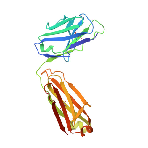
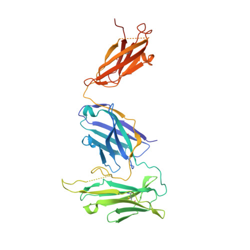
The structure of a LAIR1-containing human antibody reveals a novel mechanism of antigen recognition.

Hsieh, F.L.Higgins, M.K.

(2017) Elife 6

  • DOI: https://doi.org/10.7554/eLife.27311
  • Primary Citation Related Structures: 
    5NST

  • PubMed Abstract: 

    Antibodies are critical components of the human adaptive immune system, providing versatile scaffolds to display diverse antigen-binding surfaces. Nevertheless, most antibodies have similar architectures, with the variable immunoglobulin domains of the heavy and light chain each providing three hypervariable loops, which are varied to generate diversity. The recent identification of a novel class of antibody in humans from malaria endemic regions of Africa was therefore surprising as one hypervariable loop contains the entire collagen-binding domain of human LAIR1. Here, we present the structure of the Fab fragment of such an antibody. We show that its antigen-binding site has adopted an architecture that positions LAIR1, while itself being occluded. This therefore represents a novel means of antigen recognition, in which the Fab fragment of an antibody acts as an adaptor, linking a human protein insert with antigen-binding potential to the constant antibody regions which mediate immune cell recruitment.


  • Organizational Affiliation
    • Department of Biochemistry, University of Oxford, Oxford, United Kingdom.

Macromolecule Content 

  • Total Structure Weight: 126.41 kDa 
  • Atom Count: 8,361 
  • Modeled Residue Count: 1,076 
  • Deposited Residue Count: 1,148 
  • Unique protein chains: 2

Macromolecules

Find similar proteins by:|  3D Structure
Entity ID: 1
MoleculeChains  Sequence LengthOrganismDetailsImage
Light Chain of antibody MGD21
A, C
214Homo sapiensMutation(s): 0 
Find similar proteins by:|  3D Structure
Entity ID: 2
MoleculeChains  Sequence LengthOrganismDetailsImage
Heavy Chain of antibody MGD21
B, D
360Homo sapiensMutation(s): 0 

Experimental Data & Validation

Experimental Data

  • Method: X-RAY DIFFRACTION
  • Resolution: 2.52 Å
  • R-Value Free:  0.267 (Depositor), 0.278 (DCC) 
  • R-Value Work:  0.219 (Depositor), 0.228 (DCC) 
  • R-Value Observed: 0.221 (Depositor) 
Space Group: C 1 2 1
Unit Cell:
Length ( Å )Angle ( ˚ )
a = 169.823α = 90
b = 86.462β = 126.65
c = 103.998γ = 90
Software Package:
Software NamePurpose
BUSTERrefinement
XDSdata reduction
XDSdata scaling
PHASERphasing

Structure Validation

View Full Validation Report


Ligand Structure Quality Assessment 


Entry History 

& Funding Information

Deposition Data


Funding OrganizationLocationGrant Number
Wellcome TrustUnited Kingdom101020/Z/13/Z

Revision History  (Full details and data files)

  • Version 1.0: 2017-10-04
    Type: Initial release
  • Version 1.1: 2019-10-16
    Changes: Data collection
  • Version 1.2: 2020-07-29
    Type: Remediation
    Reason: Carbohydrate remediation
    Changes: Data collection, Derived calculations, Structure summary
  • Version 1.3: 2024-01-17
    Changes: Data collection, Database references, Refinement description, Structure summary
  • Version 1.4: 2024-11-20
    Changes: Structure summary