5NG1 | pdb_00005ng1

TUBULIN-MTC-zampanolide complex


Experimental Data Snapshot

  • Method: X-RAY DIFFRACTION
  • Resolution: 2.20 Å
  • R-Value Free: 
    0.220 (Depositor), 0.222 (DCC) 
  • R-Value Work: 
    0.173 (Depositor), 0.176 (DCC) 
  • R-Value Observed: 
    0.175 (Depositor) 

Starting Model: experimental
View more details

wwPDB Validation 3D Report Full Report

Validation slider image for 5NG1

Ligand Structure Quality Assessment 


This is version 1.4 of the entry. See complete history

Literature

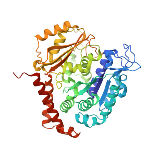
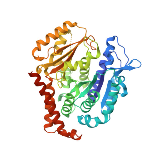
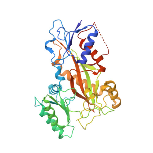
Zampanolide Binding to Tubulin Indicates Cross-Talk of Taxane Site with Colchicine and Nucleotide Sites.

Field, J.J.Pera, B.Gallego, J.E.Calvo, E.Rodriguez-Salarichs, J.Saez-Calvo, G.Zuwerra, D.Jordi, M.Andreu, J.M.Prota, A.E.Menchon, G.Miller, J.H.Altmann, K.H.Diaz, J.F.

(2018) J Nat Prod 81: 494-505

  • DOI: https://doi.org/10.1021/acs.jnatprod.7b00704
  • Primary Citation Related Structures: 
    5NFZ, 5NG1

  • PubMed Abstract: 

    The marine natural product zampanolide and analogues thereof constitute a new chemotype of taxoid site microtubule-stabilizing agents with a covalent mechanism of action. Zampanolide-ligated tubulin has the switch-activation loop (M-loop) in the assembly prone form and, thus, represents an assembly activated state of the protein. In this study, we have characterized the biochemical properties of the covalently modified, activated tubulin dimer, and we have determined the effect of zampanolide on tubulin association and the binding of tubulin ligands at other binding sites. Tubulin activation by zampanolide does not affect its longitudinal oligomerization but does alter its lateral association properties. The covalent binding of zampanolide to β-tubulin affects both the colchicine site, causing a change of the quantum yield of the bound ligand, and the exchangeable nucleotide binding site, reducing the affinity for the nucleotide. While these global effects do not change the binding affinity of 2-methoxy-5-(2,3,4-trimethoxyphenyl)-2,4,6-cycloheptatrien-1-one (MTC) (a reversible binder of the colchicine site), the binding affinity of a fluorescent analogue of GTP (Mant-GTP) at the nucleotide E-site is reduced from 12 ± 2 × 10 5 M -1 in the case of unmodified tubulin to 1.4 ± 0.3 × 10 5 M -1 in the case of the zampanolide tubulin adduct, indicating signal transmission between the taxane site and the colchicine and nucleotide sites of β-tubulin.


  • Organizational Affiliation
    • Centre for Biodiscovery, School of Biological Sciences , Victoria University of Wellington , Wellington 6012 , New Zealand.

Macromolecule Content 

  • Total Structure Weight: 266.16 kDa 
  • Atom Count: 18,369 
  • Modeled Residue Count: 2,207 
  • Deposited Residue Count: 2,319 
  • Unique protein chains: 4

Macromolecules

Find similar proteins by:|  3D Structure
Entity ID: 1
MoleculeChains  Sequence LengthOrganismDetailsImage
Tubulin alpha-1B chain
A, C
451Bos taurusMutation(s): 0 
EC: 3.6.5
UniProt
Find proteins for P81947 (Bos taurus)
Explore P81947 
Go to UniProtKB:  P81947
Entity Groups
Sequence Clusters30% Identity50% Identity70% Identity90% Identity95% Identity100% Identity
UniProt GroupP81947
Sequence Annotations
Expand
Reference Sequence
Find similar proteins by:|  3D Structure
Entity ID: 2
MoleculeChains  Sequence LengthOrganismDetailsImage
Tubulin beta-2B chain
B, D
445Bos taurusMutation(s): 0 
UniProt
Find proteins for Q6B856 (Bos taurus)
Explore Q6B856 
Go to UniProtKB:  Q6B856
Entity Groups
Sequence Clusters30% Identity50% Identity70% Identity90% Identity95% Identity100% Identity
UniProt GroupQ6B856
Sequence Annotations
Expand
Reference Sequence
Find similar proteins by:|  3D Structure
Entity ID: 3
MoleculeChains  Sequence LengthOrganismDetailsImage
Stathmin-4143Rattus norvegicusMutation(s): 0 
Gene Names: Stmn4
UniProt
Find proteins for P63043 (Rattus norvegicus)
Explore P63043 
Go to UniProtKB:  P63043
Entity Groups
Sequence Clusters30% Identity50% Identity70% Identity90% Identity95% Identity100% Identity
UniProt GroupP63043
Sequence Annotations
Expand
Reference Sequence
Find similar proteins by:|  3D Structure
Entity ID: 4
MoleculeChains  Sequence LengthOrganismDetailsImage
Tubulin-Tyrosine Ligase384Gallus gallusMutation(s): 0 
Gene Names: TTL

Small Molecules

Ligands 10 Unique
IDChains Name / Formula / InChI Key2D Diagram3D Interactions
GTP

Query on GTP


Download:Ideal Coordinates CCD File
G [auth A],
P [auth C]
GUANOSINE-5'-TRIPHOSPHATE
C10 H16 N5 O14 P3
XKMLYUALXHKNFT-UUOKFMHZSA-N
ACP

Query on ACP


Download:Ideal Coordinates CCD File
W [auth F]PHOSPHOMETHYLPHOSPHONIC ACID ADENYLATE ESTER
C11 H18 N5 O12 P3
UFZTZBNSLXELAL-IOSLPCCCSA-N
8WE

Query on 8WE


Download:Ideal Coordinates CCD File
M [auth B](2~{Z},4~{E})-~{N}-[(~{S})-oxidanyl-[(1~{S},2~{E},5~{S},11~{R},17~{S},19~{R})-3,11,19-trimethyl-7,13-bis(oxidanylidene)-6,21-dioxabicyclo[15.3.1]henicos-2-en-5-yl]methyl]hexa-2,4-dienamide
C29 H45 N O6
WSHDEXCLDGAKJW-NFPHSYLMSA-N
ZPN

Query on ZPN


Download:Ideal Coordinates CCD File
V [auth D](2Z,4E)-N-[(S)-[(1S,2E,5S,8E,10Z,17S)-3,11-dimethyl-19-methylidene-7,13-dioxo-6,21-dioxabicyclo[15.3.1]henicosa-2,8,10-trien-5-yl](hydroxy)methyl]hexa-2,4-dienamide
C29 H39 N O6
KNEMNZWQWFRUIB-KZVIXHMTSA-N
GDP

Query on GDP


Download:Ideal Coordinates CCD File
J [auth B],
S [auth D]
GUANOSINE-5'-DIPHOSPHATE
C10 H15 N5 O11 P2
QGWNDRXFNXRZMB-UUOKFMHZSA-N
8WB

Query on 8WB


Download:Ideal Coordinates CCD File
L [auth B],
U [auth D]
2-methoxy-5-(2,3,4-trimethoxyphenyl)cyclohepta-2,4,6-trien-1-one
C17 H18 O5
QIMGSZURBOTPMW-UHFFFAOYSA-N
MES

Query on MES


Download:Ideal Coordinates CCD File
O [auth B]2-(N-MORPHOLINO)-ETHANESULFONIC ACID
C6 H13 N O4 S
SXGZJKUKBWWHRA-UHFFFAOYSA-N
GOL

Query on GOL


Download:Ideal Coordinates CCD File
N [auth B]GLYCEROL
C3 H8 O3
PEDCQBHIVMGVHV-UHFFFAOYSA-N
CA

Query on CA


Download:Ideal Coordinates CCD File
I [auth A],
R [auth C]
CALCIUM ION
Ca
BHPQYMZQTOCNFJ-UHFFFAOYSA-N
MG

Query on MG


Download:Ideal Coordinates CCD File
H [auth A],
K [auth B],
Q [auth C],
T [auth D],
X [auth F]
MAGNESIUM ION
Mg
JLVVSXFLKOJNIY-UHFFFAOYSA-N

Experimental Data & Validation

Experimental Data

  • Method: X-RAY DIFFRACTION
  • Resolution: 2.20 Å
  • R-Value Free:  0.220 (Depositor), 0.222 (DCC) 
  • R-Value Work:  0.173 (Depositor), 0.176 (DCC) 
  • R-Value Observed: 0.175 (Depositor) 
Space Group: P 21 21 21
Unit Cell:
Length ( Å )Angle ( ˚ )
a = 104.237α = 90
b = 157.418β = 90
c = 178.858γ = 90
Software Package:
Software NamePurpose
PHENIXrefinement
XDSdata reduction
XSCALEdata scaling
PHENIXphasing

Structure Validation

View Full Validation Report


Ligand Structure Quality Assessment 


Entry History 

Revision History  (Full details and data files)

  • Version 1.0: 2017-10-25
    Type: Initial release
  • Version 1.1: 2018-04-04
    Changes: Data collection, Database references
  • Version 1.2: 2018-05-02
    Changes: Data collection
  • Version 1.3: 2024-01-17
    Changes: Data collection, Database references, Derived calculations, Refinement description
  • Version 1.4: 2024-10-16
    Changes: Structure summary