5NF0 | pdb_00005nf0

Discovery, crystal structures and atomic force microscopy study of thioether ligated D,L-cyclic antimicrobial peptides against multidrug resistant Pseudomonas aeruginosa


Experimental Data Snapshot

  • Method: X-RAY DIFFRACTION
  • Resolution: 1.27 Å
  • R-Value Free: 
    0.184 (Depositor), 0.185 (DCC) 
  • R-Value Work: 
    0.163 (Depositor), 0.163 (DCC) 
  • R-Value Observed: 
    0.164 (Depositor) 

Starting Model: experimental
View more details

wwPDB Validation 3D Report Full Report

Validation slider image for 5NF0

Ligand Structure Quality Assessment 


This is version 2.0 of the entry. See complete history

Literature

Design, crystal structure and atomic force microscopy study of thioether ligated d,l-cyclic antimicrobial peptides against multidrug resistant Pseudomonas aeruginosa.

He, R.Di Bonaventura, I.Visini, R.Gan, B.H.Fu, Y.Probst, D.Luscher, A.Kohler, T.van Delden, C.Stocker, A.Hong, W.Darbre, T.Reymond, J.L.

(2017) Chem Sci 8: 7464-7475

  • DOI: https://doi.org/10.1039/c7sc01599b
  • Primary Citation Related Structures: 
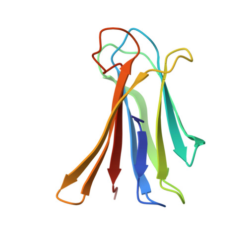
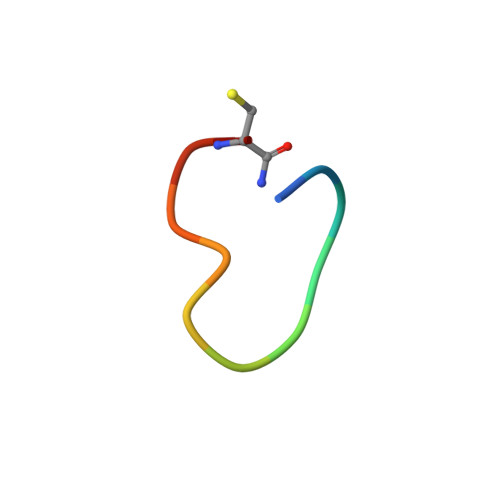
    5NES, 5NEY, 5NF0

  • PubMed Abstract: 

    Here we report a new family of cyclic antimicrobial peptides (CAMPs) targeting MDR strains of Pseudomonas aeruginosa . These CAMPs are cyclized via a xylene double thioether bridge connecting two cysteines placed at the ends of a linear amphiphilic alternating d,l-sequence composed of lysines and tryptophans. Investigations by transmission electron microscopy (TEM), dynamic light scattering and atomic force microscopy (AFM) suggest that these peptide macrocycles interact with the membrane to form lipid-peptide aggregates. Amphiphilic conformations compatible with membrane disruption are observed in high resolution X-ray crystal structures of fucosylated derivatives in complex with lectin LecB. The potential for optimization is highlighted by N -methylation of backbone amides leading to derivatives with similar antimicrobial activity but lower hemolysis.


  • Organizational Affiliation
    • Department of Chemistry and Biochemistry , University of Bern , Freiestrasse 3 , 3012 Bern , Switzerland . Email: jean-louis.reymond@dcb.unibe.ch.

Macromolecule Content 

  • Total Structure Weight: 53.54 kDa 
  • Atom Count: 4,303 
  • Modeled Residue Count: 493 
  • Deposited Residue Count: 497 
  • Unique protein chains: 3

Macromolecules

Find similar proteins by:|  3D Structure
Entity ID: 1
MoleculeChains  Sequence LengthOrganismDetailsImage
Fucose-binding lectin II (PA-IIL)
A, B, C, D
114Pseudomonas aeruginosaMutation(s): 0 
Gene Names: lecBPAERUG_E15_London_28_01_14_00983PAERUG_P32_London_17_VIM_2_10_11_00423PAMH19_1713
UniProt
Find proteins for Q9HYN5 (Pseudomonas aeruginosa (strain ATCC 15692 / DSM 22644 / CIP 104116 / JCM 14847 / LMG 12228 / 1C / PRS 101 / PAO1))
Explore Q9HYN5 
Go to UniProtKB:  Q9HYN5
Entity Groups
Sequence Clusters30% Identity50% Identity70% Identity90% Identity95% Identity100% Identity
UniProt GroupQ9HYN5
Sequence Annotations
Expand
Reference Sequence
Find similar proteins by:  Sequence   |   3D Structure  
Entity ID: 2
MoleculeChains  Sequence LengthOrganismDetailsImage
CYD-TRP-TRD-LYS-LYD-LYS-LYD-LYS-TRD-TRP-CYD-GLY
E, G, H
12synthetic constructMutation(s): 0 
Find similar proteins by:  Sequence   |   3D Structure  
Entity ID: 3
MoleculeChains  Sequence LengthOrganismDetailsImage
Fragment of ligand5synthetic constructMutation(s): 0 

Small Molecules

Ligands 3 Unique
IDChains Name / Formula / InChI Key2D Diagram3D Interactions
ZDC

Query on ZDC


Download:Ideal Coordinates CCD File
Q [auth E],
S [auth F],
T [auth G],
V [auth H]
3,7-anhydro-2,8-dideoxy-L-glycero-D-gluco-octonic acid
C8 H14 O6
YTZUDUWVQZSKNN-OASCRQMUSA-N
OXE

Query on OXE


Download:Ideal Coordinates CCD File
R [auth E],
U [auth G],
W [auth H]
ORTHO-XYLENE
C8 H10
CTQNGGLPUBDAKN-UHFFFAOYSA-N
CA

Query on CA


Download:Ideal Coordinates CCD File
I [auth A]
J [auth A]
K [auth B]
L [auth B]
M [auth C]
I [auth A],
J [auth A],
K [auth B],
L [auth B],
M [auth C],
N [auth C],
O [auth D],
P [auth D]
CALCIUM ION
Ca
BHPQYMZQTOCNFJ-UHFFFAOYSA-N

Experimental Data & Validation

Experimental Data

  • Method: X-RAY DIFFRACTION
  • Resolution: 1.27 Å
  • R-Value Free:  0.184 (Depositor), 0.185 (DCC) 
  • R-Value Work:  0.163 (Depositor), 0.163 (DCC) 
  • R-Value Observed: 0.164 (Depositor) 
Space Group: P 21 21 21
Unit Cell:
Length ( Å )Angle ( ˚ )
a = 45.278α = 90
b = 99.966β = 90
c = 106.611γ = 90
Software Package:
Software NamePurpose
PHENIXrefinement
XDSdata reduction
XDSdata scaling
PHENIXphasing

Structure Validation

View Full Validation Report


Ligand Structure Quality Assessment 


Entry History 

Deposition Data

Revision History  (Full details and data files)

  • Version 1.0: 2017-09-13
    Type: Initial release
  • Version 1.1: 2017-12-06
    Changes: Database references, Derived calculations
  • Version 1.2: 2020-07-29
    Type: Remediation
    Reason: Carbohydrate remediation
    Changes: Data collection, Derived calculations, Refinement description
  • Version 1.3: 2024-01-17
    Changes: Data collection, Database references, Refinement description
  • Version 2.0: 2025-10-01
    Changes: Advisory, Atomic model, Data collection, Derived calculations, Other, Structure summary