Conformational Analysis of the Mannosidase Inhibitor Kifunensine: A Quantum Mechanical and Structural Approach.
Males, A., Raich, L., Williams, S.J., Rovira, C., Davies, G.J.(2017) Chembiochem 18: 1496-1501
- PubMed: 28493500 Search on PubMed
- DOI: https://doi.org/10.1002/cbic.201700166
- Primary Citation Related Structures:
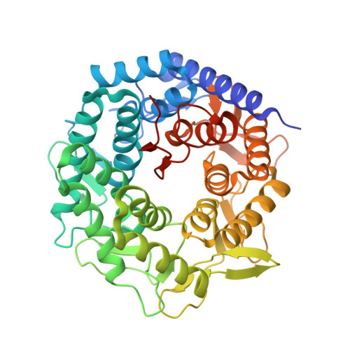
5NE5 - PubMed Abstract:
The varied yet family-specific conformational pathways used by individual glycoside hydrolases (GHs) offer a tantalising prospect for the design of tightly binding and specific enzyme inhibitors. A cardinal example of a GH-family-specific inhibitor, and one that finds widespread practical use, is the natural product kifunensine, which is a low-nanomolar inhibitor that is selective for GH family 47 inverting α-mannosidases. Here we show, through quantum-mechanical approaches, that kifunensine is restrained to a "ring-flipped" 1 C 4 conformation with another accessible, but higher-energy, region around the 1,4 B conformation. The conformations of kifunensine in complex with a range of GH47 enzymes-including an atomic-level resolution (1 Å) structure of kifunensine with Caulobacter sp. CkGH47 reported herein and with GH family 38 and 92 α-mannosidases-were mapped onto the kifunensine free-energy landscape. These studies revealed that kifunensine has the ability to mimic the product state of GH47 enzymes but cannot mimic any conformational states relevant to the reaction coordinate of mannosidases from other families.
- York Structural Biology Laboratory, Department of Chemistry, The University of York, York, YO10 5DD, UK.
Organizational Affiliation: