5N7K | pdb_00005n7k

Crystal structure of the coiled-coil domain of human tricellulin


Experimental Data Snapshot

  • Method: X-RAY DIFFRACTION
  • Resolution: 2.81 Å
  • R-Value Free: 
    0.242 (Depositor), 0.252 (DCC) 
  • R-Value Work: 
    0.215 (Depositor), 0.220 (DCC) 
  • R-Value Observed: 
    0.217 (Depositor) 

Starting Model: experimental
View more details

wwPDB Validation 3D Report Full Report

Validation slider image for 5N7K

This is version 1.2 of the entry. See complete history

Literature

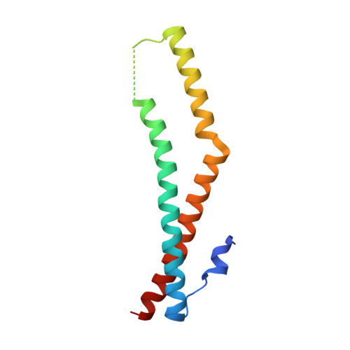
Crystal structure of the tricellulin C-terminal coiled-coil domain reveals a unique mode of dimerization.

Schuetz, A.Radusheva, V.Krug, S.M.Heinemann, U.

(2017) Ann N Y Acad Sci 1405: 147-159

  • DOI: https://doi.org/10.1111/nyas.13408
  • Primary Citation Related Structures: 
    5N7H, 5N7I, 5N7K

  • PubMed Abstract: 

    Tricellulin is a tight junction protein localized to tricellular contacts in many epithelial tissues, where it is required for full barrier control. Here, we present crystal structures of the tricellulin C-terminal coiled-coil domain, revealing a potential dimeric arrangement. By combining structural, biochemical, functional, and mutation analyses, we gain insight into the mode of tricellulin oligomerization and suggest a model where dimerization of its cytoplasmic C-terminus may play an auxiliary role in stabilizing homophilic and potentially also heterophilic cis-interactions within tight junctions.


  • Organizational Affiliation
    • Helmholtz Protein Sample Production Facility, Max Delbrück Center for Molecular Medicine, Berlin, Germany.

Macromolecule Content 

  • Total Structure Weight: 55.91 kDa 
  • Atom Count: 3,789 
  • Modeled Residue Count: 442 
  • Deposited Residue Count: 460 
  • Unique protein chains: 1

Macromolecules

Find similar proteins by:|  3D Structure
Entity ID: 1
MoleculeChains  Sequence LengthOrganismDetailsImage
MARVEL domain-containing protein 2
A, B, C, D
115Homo sapiensMutation(s): 0 
Gene Names: MARVELD2TRIC
UniProt & NIH Common Fund Data Resources
Find proteins for Q8N4S9 (Homo sapiens)
Explore Q8N4S9 
Go to UniProtKB:  Q8N4S9
PHAROS:  Q8N4S9
GTEx:  ENSG00000152939 
Entity Groups
Sequence Clusters30% Identity50% Identity70% Identity90% Identity95% Identity100% Identity
UniProt GroupQ8N4S9
Sequence Annotations
Expand
Reference Sequence

Experimental Data & Validation

Experimental Data

  • Method: X-RAY DIFFRACTION
  • Resolution: 2.81 Å
  • R-Value Free:  0.242 (Depositor), 0.252 (DCC) 
  • R-Value Work:  0.215 (Depositor), 0.220 (DCC) 
  • R-Value Observed: 0.217 (Depositor) 
Space Group: P 32 2 1
Unit Cell:
Length ( Å )Angle ( ˚ )
a = 82.621α = 90
b = 82.621β = 90
c = 188.825γ = 120
Software Package:
Software NamePurpose
BUSTERrefinement
XDSdata reduction
XDSdata scaling
PHASERphasing

Structure Validation

View Full Validation Report


Entry History 

Deposition Data

Revision History  (Full details and data files)

  • Version 1.0: 2017-07-12
    Type: Initial release
  • Version 1.1: 2017-10-18
    Changes: Database references
  • Version 1.2: 2024-01-17
    Changes: Data collection, Database references, Refinement description