5N4S

VIM-2 metallo-beta-lactamase in complex with ((S)-3-mercapto-2-methylpropanoyl)-D-tryptophan (Compound 3)


Experimental Data Snapshot

  • Method: X-RAY DIFFRACTION
  • Resolution: 1.20 Å
  • R-Value Free: 0.200 
  • R-Value Work: 0.184 
  • R-Value Observed: 0.185 

wwPDB Validation   3D Report Full Report


Ligand Structure Quality Assessment 


This is version 1.3 of the entry. See complete history


Literature

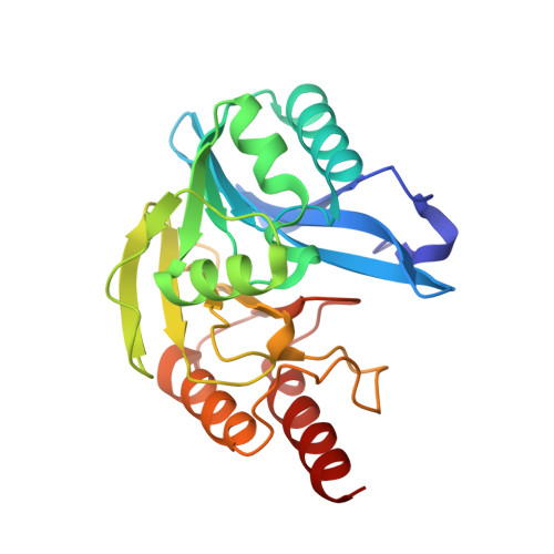
Crystallographic analyses of isoquinoline complexes reveal a new mode of metallo-beta-lactamase inhibition.

Li, G.B.Brem, J.Lesniak, R.Abboud, M.I.Lohans, C.T.Clifton, I.J.Yang, S.Y.Jimenez-Castellanos, J.C.Avison, M.B.Spencer, J.McDonough, M.A.Schofield, C.J.

(2017) Chem Commun (Camb) 53: 5806-5809

  • DOI: https://doi.org/10.1039/c7cc02394d
  • Primary Citation of Related Structures:  
    5N4S, 5N4T, 5N55, 5N58, 5NAI

  • PubMed Abstract: 

    Crystallographic analyses of the VIM-5 metallo-β-lactamase (MBL) with isoquinoline inhibitors reveal non zinc ion binding modes. Comparison with other MBL-inhibitor structures directed addition of a zinc-binding thiol enabling identification of potent B1 MBL inhibitors. The inhibitors potentiate meropenem activity against clinical isolates harboring MBLs.


  • Organizational Affiliation

    Department of Chemistry, University of Oxford, 12 Mansfield Road, Oxford, OX1 3TA, UK. christopher.schofield@chem.ox.ac.uk jurgen.brem@chem.ox.ac.uk.


Macromolecules
Find similar proteins by:  (by identity cutoff)  |  3D Structure
Entity ID: 1
MoleculeChains Sequence LengthOrganismDetailsImage
Beta-lactamase VIM-2
A, B
232Pseudomonas aeruginosaMutation(s): 0 
Gene Names: blaVIM-2bla vim-2bla-VIM-2blasVIM-2blaVIM2blmVIM-2PAERUG_P32_London_17_VIM_2_10_11_06255
UniProt
Find proteins for Q9K2N0 (Pseudomonas aeruginosa)
Explore Q9K2N0 
Go to UniProtKB:  Q9K2N0
Entity Groups  
Sequence Clusters30% Identity50% Identity70% Identity90% Identity95% Identity100% Identity
UniProt GroupQ9K2N0
Sequence Annotations
Expand
  • Reference Sequence
Small Molecules
Ligands 3 Unique
IDChains Name / Formula / InChI Key2D Diagram3D Interactions
R38
Query on R38

Download Ideal Coordinates CCD File 
F [auth A],
L [auth B]
(2~{R})-3-(1~{H}-indol-3-yl)-2-[[(2~{S})-2-methyl-3-sulfanyl-propanoyl]amino]propanoic acid
C15 H18 N2 O3 S
ZOUTYVWHWSUKPL-NOZJJQNGSA-N
ZN
Query on ZN

Download Ideal Coordinates CCD File 
C [auth A]
D [auth A]
E [auth A]
I [auth B]
J [auth B]
C [auth A],
D [auth A],
E [auth A],
I [auth B],
J [auth B],
K [auth B]
ZINC ION
Zn
PTFCDOFLOPIGGS-UHFFFAOYSA-N
FMT
Query on FMT

Download Ideal Coordinates CCD File 
G [auth A]
H [auth A]
M [auth B]
N [auth B]
O [auth B]
G [auth A],
H [auth A],
M [auth B],
N [auth B],
O [auth B],
P [auth B]
FORMIC ACID
C H2 O2
BDAGIHXWWSANSR-UHFFFAOYSA-N
Binding Affinity Annotations 
IDSourceBinding Affinity
R38 Binding MOAD:  5N4S IC50: 55 (nM) from 1 assay(s)
Experimental Data & Validation

Experimental Data

  • Method: X-RAY DIFFRACTION
  • Resolution: 1.20 Å
  • R-Value Free: 0.200 
  • R-Value Work: 0.184 
  • R-Value Observed: 0.185 
  • Space Group: C 1 2 1
Unit Cell:
Length ( Å )Angle ( ˚ )
a = 103.845α = 90
b = 80.308β = 130.34
c = 68.732γ = 90
Software Package:
Software NamePurpose
PHENIXrefinement
SCALEPACKdata scaling
PDB_EXTRACTdata extraction
PHASERphasing
HKL-3000data reduction

Structure Validation

View Full Validation Report


Ligand Structure Quality Assessment 


Entry History 

Deposition Data

Revision History  (Full details and data files)

  • Version 1.0: 2017-05-17
    Type: Initial release
  • Version 1.1: 2017-06-07
    Changes: Database references
  • Version 1.2: 2019-10-16
    Changes: Data collection
  • Version 1.3: 2024-01-17
    Changes: Data collection, Database references, Derived calculations, Refinement description