5MX7 | pdb_00005mx7

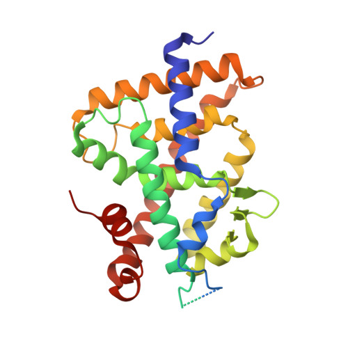
1a,20S-dihydroxyvitamin D3 VDR complex


Experimental Data Snapshot

  • Method: X-RAY DIFFRACTION
  • Resolution: 1.98 Å
  • R-Value Free: 
    0.225 (Depositor), 0.216 (DCC) 
  • R-Value Work: 
    0.200 (Depositor) 
  • R-Value Observed: 
    0.201 (Depositor) 

Starting Model: experimental
View more details

wwPDB Validation 3D Report Full Report

Validation slider image for 5MX7

Ligand Structure Quality Assessment 


This is version 1.2 of the entry. See complete history. 

Literature

1 alpha,20S-Dihydroxyvitamin D3 Interacts with Vitamin D Receptor: Crystal Structure and Route of Chemical Synthesis.

Lin, Z., Chen, H., Belorusova, A.Y., Bollinger, J.C., Tang, E.K.Y., Janjetovic, Z., Kim, T.K., Wu, Z., Miller, D.D., Slominski, A.T., Postlethwaite, A.E., Tuckey, R.C., Rochel, N., Li, W.

(2017) Sci Rep 7: 10193-10193

  • DOI: https://doi.org/10.1038/s41598-017-10917-7
  • Primary Citation Related Structures: 
    5MX7

  • PubMed Abstract: 

    1α,20S-Dihydroxyvitamin D3 [1,20S(OH) 2 D 3 ], a natural and bioactive vitamin D3 metabolite, was chemically synthesized for the first time. X-ray crystallography analysis of intermediate 15 confirmed its 1α-OH configuration. 1,20S(OH) 2 D 3 interacts with the vitamin D receptor (VDR), with similar potency to its native ligand, 1α,25-dihydroxyvitamin D 3 [1,25(OH) 2 D 3 ] as illustrated by its ability to stimulate translocation of the VDR to the nucleus, stimulate VDRE-reporter activity, regulate VDR downstream genes (VDR, CYP24A1, TRPV6 and CYP27B1), and inhibit the production of inflammatory markers (IFNγ and IL1β). However, their co-crystal structures revealed differential molecular interactions of the 20S-OH moiety and the 25-OH moiety to the VDR, which may explain some differences in their biological activities. Furthermore, this study provides a synthetic route for the synthesis of 1,20S(OH) 2 D 3 using the intermediate 1α,3β-diacetoxypregn-5-en-20-one (3), and provides a molecular and biological basis for the development of 1,20S(OH) 2 D 3 and its analogs as potential therapeutic agents.


  • Organizational Affiliation: 
    • Department of Pharmaceutical Sciences, University of Tennessee Health Science Center, 881 Madison Avenue, Room 561, Memphis, TN, 38163, United States.

Macromolecule Content 

  • Total Structure Weight: 36.11 kDa 
  • Atom Count: 2,080 
  • Modeled Residue Count: 248 
  • Deposited Residue Count: 315 
  • Unique protein chains: 2

Macromolecules

Find similar proteins by:|  3D Structure
Entity ID: 1
MoleculeChains  Sequence LengthOrganismDetailsImage
Vitamin D3 receptor AA [auth A1]300Danio rerioMutation(s): 0 
Gene Names: vdra, nr1i1a, vdr
UniProt
Find proteins for Q9PTN2 (Danio rerio)
Explore Q9PTN2 
Go to UniProtKB:  Q9PTN2
Entity Groups
Sequence Clusters30% Identity50% Identity70% Identity90% Identity95% Identity100% Identity
UniProt GroupQ9PTN2
Sequence Annotations
Expand
Reference Sequence
Find similar proteins by:  Sequence   |   3D Structure  
Entity ID: 2
MoleculeChains  Sequence LengthOrganismDetailsImage
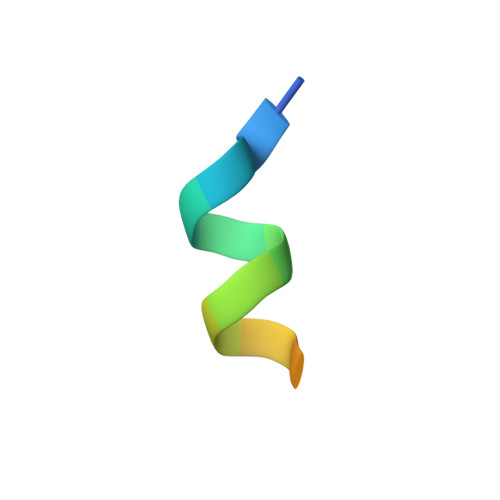
Nuclear receptor coactivator 1B [auth B1]15Homo sapiensMutation(s): 0 
EC: 2.3.1.48
UniProt & NIH Common Fund Data Resources
Find proteins for Q15788 (Homo sapiens)
Explore Q15788 
Go to UniProtKB:  Q15788
PHAROS:  Q15788
GTEx:  ENSG00000084676 
Entity Groups
Sequence Clusters30% Identity50% Identity70% Identity90% Identity95% Identity100% Identity
UniProt GroupQ15788
Sequence Annotations
Expand
Reference Sequence

Small Molecules

Ligands 1 Unique
IDChains Name / Formula / InChI Key2D Diagram3D Interactions
D3V

Query on D3V


Download:Ideal Coordinates CCD File
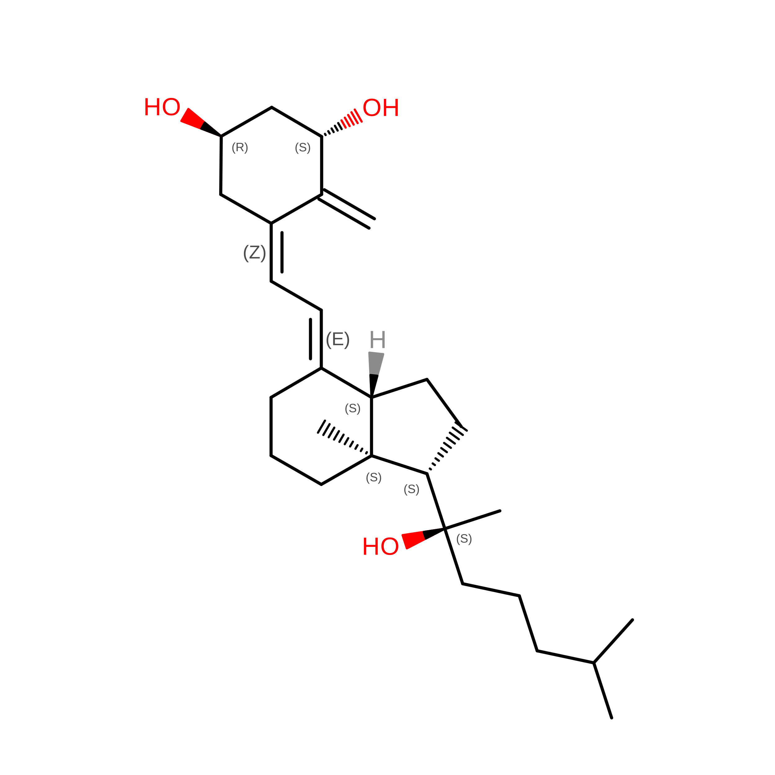
C [auth A1] 1a,20S-dihydroxyvitamin D3
C27 H44 O3
RLDKSDBKVGPMKA-YAUUYYILSA-N

Experimental Data & Validation

Experimental Data

  • Method: X-RAY DIFFRACTION
  • Resolution: 1.98 Å
  • R-Value Free:  0.225 (Depositor), 0.216 (DCC) 
  • R-Value Work:  0.200 (Depositor) 
  • R-Value Observed: 0.201 (Depositor) 
Space Group: P 65 2 2
Unit Cell:
Length ( Å )Angle ( ˚ )
a = 65.936α = 90
b = 65.936β = 90
c = 262.97γ = 120
Software Package:
Software NamePurpose
PHENIXrefinement
HKL-2000data reduction
HKL-2000data scaling
PHENIXphasing

Structure Validation

View Full Validation Report


Ligand Structure Quality Assessment 


Entry History 

& Funding Information

Deposition Data


Funding OrganizationLocationGrant Number
French National Research AgencyFranceANR-13-BSV8-0024-01

Revision History  (Full details and data files)

  • Version 1.0: 2017-11-29
    Type: Initial release
  • Version 1.1: 2021-02-24
    Changes: Derived calculations
  • Version 1.2: 2024-01-17
    Changes: Data collection, Database references, Refinement description