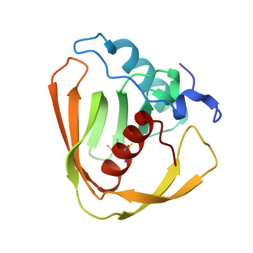
5MTE

Crystal structure of PDF from the Vibrio parahaemolyticus bacteriophage VP16T in complex with actinonin - crystal form II


Experimental Data Snapshot

  • Method: X-RAY DIFFRACTION
  • Resolution: 1.40 Å
  • R-Value Free: 0.203 
  • R-Value Work: 0.187 
  • R-Value Observed: 0.188 

Starting Model: experimental
View more details

wwPDB Validation   3D Report Full Report


Ligand Structure Quality Assessment 


This is version 1.3 of the entry. See complete history


Literature

Peptide deformylases from Vibrio parahaemolyticus phage and bacteria display similar deformylase activity and inhibitor binding clefts.

Grzela, R.Nusbaum, J.Fieulaine, S.Lavecchia, F.Desmadril, M.Nhiri, N.Van Dorsselaer, A.Cianferani, S.Jacquet, E.Meinnel, T.Giglione, C.

(2018) Biochim Biophys Acta 1866: 348-355

  • DOI: https://doi.org/10.1016/j.bbapap.2017.10.007
  • Primary Citation of Related Structures:  
    5MTE

  • PubMed Abstract: 

    Unexpected peptide deformylase (PDF) genes were recently retrieved in numerous marine phage genomes. While various hypotheses dealing with the occurrence of these intriguing sequences have been made, no further characterization and functional studies have been described thus far. In this study, we characterize the bacteriophage Vp16 PDF enzyme, as representative member of the newly identified C-terminally truncated viral PDFs. We show here that conditions classically used for bacterial PDFs lead to an enzyme exhibiting weak activity. Nonetheless, our integrated biophysical and biochemical approaches reveal specific effects of pH and metals on Vp16 PDF stability and activity. A novel purification protocol taking in account these data allowed strong improvement of Vp16 PDF specific activity to values similar to those of bacterial PDFs. We next show that Vp16 PDF is as sensitive to the natural inhibitor compound of PDFs, actinonin, as bacterial PDFs. Comparison of the 3D structures of Vp16 and E. coli PDFs bound to actinonin also reveals that both PDFs display identical substrate binding mode. We conclude that bacteriophage Vp16 PDF protein has functional peptide deformylase activity and we suggest that encoded phage PDFs might be important for viral fitness.


  • Organizational Affiliation

    Institute for Integrative Biology of the Cell (I2BC), CEA, CNRS, Univ. Paris-Sud, Université Paris-Saclay, 91198 Gif-sur-Yvette Cedex, France.


Macromolecules
Find similar proteins by:  (by identity cutoff)  |  3D Structure
Entity ID: 1
MoleculeChains Sequence LengthOrganismDetailsImage
Putative uncharacterized protein orf60T
A, B
137Vibrio phage VP16TMutation(s): 0 
Gene Names: orf60T
UniProt
Find proteins for Q6VT21 (Vibrio phage VP16T)
Explore Q6VT21 
Go to UniProtKB:  Q6VT21
Entity Groups  
Sequence Clusters30% Identity50% Identity70% Identity90% Identity95% Identity100% Identity
UniProt GroupQ6VT21
Sequence Annotations
Expand
  • Reference Sequence
Small Molecules
Ligands 3 Unique
IDChains Name / Formula / InChI Key2D Diagram3D Interactions
BB2
Query on BB2

Download Ideal Coordinates CCD File 
C [auth A],
F [auth B]
ACTINONIN
C19 H35 N3 O5
XJLATMLVMSFZBN-VYDXJSESSA-N
ZN
Query on ZN

Download Ideal Coordinates CCD File 
D [auth A],
G [auth B]
ZINC ION
Zn
PTFCDOFLOPIGGS-UHFFFAOYSA-N
NI
Query on NI

Download Ideal Coordinates CCD File 
E [auth A],
H [auth B],
I [auth B]
NICKEL (II) ION
Ni
VEQPNABPJHWNSG-UHFFFAOYSA-N
Experimental Data & Validation

Experimental Data

  • Method: X-RAY DIFFRACTION
  • Resolution: 1.40 Å
  • R-Value Free: 0.203 
  • R-Value Work: 0.187 
  • R-Value Observed: 0.188 
  • Space Group: P 32 2 1
Unit Cell:
Length ( Å )Angle ( ˚ )
a = 64.62α = 90
b = 64.62β = 90
c = 124.89γ = 120
Software Package:
Software NamePurpose
REFMACrefinement
XDSdata reduction
XDSdata scaling
PHASERphasing

Structure Validation

View Full Validation Report


Ligand Structure Quality Assessment 


Entry History & Funding Information

Deposition Data


Funding OrganizationLocationGrant Number
French National Research AgencyFranceANR-10-Blanc-1510

Revision History  (Full details and data files)

  • Version 1.0: 2017-11-29
    Type: Initial release
  • Version 1.1: 2018-01-03
    Changes: Database references
  • Version 1.2: 2024-01-17
    Changes: Data collection, Database references, Derived calculations, Refinement description
  • Version 1.3: 2024-10-16
    Changes: Structure summary