5MSF | pdb_00005msf

MS2 PROTEIN CAPSID/RNA COMPLEX


Experimental Data Snapshot

  • Method: X-RAY DIFFRACTION
  • Resolution: 2.80 Å
  • R-Value Free: 
    0.197 (Depositor), 0.188 (DCC) 
  • R-Value Work: 
    0.188 (Depositor), 0.185 (DCC) 

Starting Model: experimental
View more details

wwPDB Validation 3D Report Full Report

Validation slider image for 5MSF

This is version 1.3 of the entry. See complete history

Literature

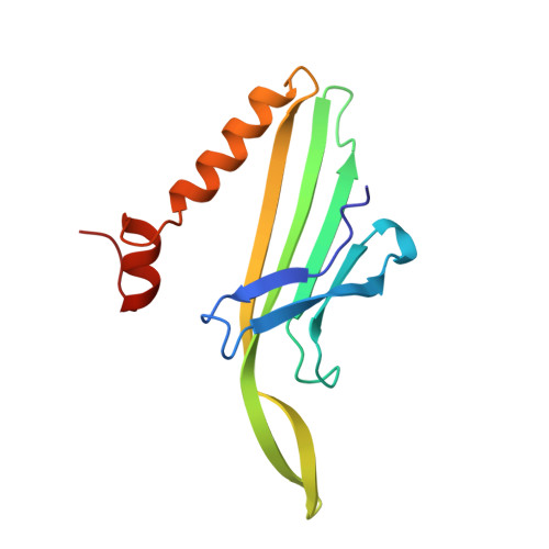
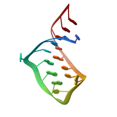
Crystal structures of a series of RNA aptamers complexed to the same protein target.

Rowsell, S.Stonehouse, N.J.Convery, M.A.Adams, C.J.Ellington, A.D.Hirao, I.Peabody, D.S.Stockley, P.G.Phillips, S.E.

(1998) Nat Struct Biol 5: 970-975

  • DOI: https://doi.org/10.1038/2946
  • Primary Citation Related Structures: 
    5MSF, 7MSF

  • PubMed Abstract: 

    We have determined the crystal structures, at 2.8 A resolution, of two different RNA aptamers, each bound to MS2 coat protein. One of the aptamers contains a non-Watson-Crick base pair, while the other is missing one of the unpaired adenines that make sequence-specific contacts in the wild-type complex. Despite these differences, the RNA aptamers bind in the same location on the protein as the wild-type translational operator. Comparison of these new structures with other MS2-RNA complexes allows us to refine further the definition of the minimal recognition elements and suggests a possible application of the MS2 system for routine structure determination of small nucleic acid motifs.


  • Organizational Affiliation
    • School of Biochemistry and Molecular Biology, and North of England Structural Biology Centre, University of Leeds, UK.

Macromolecule Content 

  • Total Structure Weight: 52.87 kDa 
  • Atom Count: 3,737 
  • Modeled Residue Count: 421 
  • Deposited Residue Count: 423 
  • Unique protein chains: 1
  • Unique nucleic acid chains: 1

Macromolecules


Find similar proteins by:|  3D Structure
Entity ID: 2
MoleculeChains  Sequence LengthOrganismDetailsImage
MS2 PROTEIN CAPSIDC [auth A],
D [auth B],
E [auth C]
129Escherichia phage MS2Mutation(s): 0 
Gene Names: COAT PROTEIN
UniProt
Find proteins for P03612 (Escherichia phage MS2)
Explore P03612 
Go to UniProtKB:  P03612
Entity Groups
Sequence Clusters30% Identity50% Identity70% Identity90% Identity95% Identity100% Identity
UniProt GroupP03612
Sequence Annotations
Expand
Reference Sequence
Find similar nucleic acids by:  Sequence
Entity ID: 1
MoleculeChains LengthOrganismImage
5'-R(*CP*CP*GP*GP*AP*GP*GP*AP*UP*CP*AP*CP*CP*AP*CP*GP*GP*G)-3'A [auth R],
B [auth S]
18N/A
Sequence Annotations
Expand
Reference Sequence

Experimental Data & Validation

Experimental Data

  • Method: X-RAY DIFFRACTION
  • Resolution: 2.80 Å
  • R-Value Free:  0.197 (Depositor), 0.188 (DCC) 
  • R-Value Work:  0.188 (Depositor), 0.185 (DCC) 
Space Group: H 3 2
Unit Cell:
Length ( Å )Angle ( ˚ )
a = 287.3α = 90
b = 287.3β = 90
c = 651.6γ = 120
Software Package:
Software NamePurpose
CCP4model building
X-PLORrefinement
MOSFLMdata reduction
CCP4data scaling
CCP4phasing

Structure Validation

View Full Validation Report


Entry History 

Deposition Data

Revision History  (Full details and data files)

  • Version 1.0: 1998-11-11
    Type: Initial release
  • Version 1.1: 2008-05-22
    Changes: Version format compliance
  • Version 1.2: 2011-07-13
    Changes: Version format compliance
  • Version 1.3: 2023-08-02
    Changes: Database references, Derived calculations, Refinement description