5MNQ

Cationic trypsin in complex with a derivative of N-amidinopiperidine


Experimental Data Snapshot

  • Method: X-RAY DIFFRACTION
  • Resolution: 1.34 Å
  • R-Value Free: 0.171 
  • R-Value Work: 0.137 
  • R-Value Observed: 0.139 

wwPDB Validation   3D Report Full Report


Ligand Structure Quality Assessment 


This is version 1.2 of the entry. See complete history


Literature

Intriguing role of water in protein-ligand binding studied by neutron crystallography on trypsin complexes.

Schiebel, J.Gaspari, R.Wulsdorf, T.Ngo, K.Sohn, C.Schrader, T.E.Cavalli, A.Ostermann, A.Heine, A.Klebe, G.

(2018) Nat Commun 9: 3559-3559

  • DOI: https://doi.org/10.1038/s41467-018-05769-2
  • Primary Citation of Related Structures:  
    5MNE, 5MNF, 5MNG, 5MNH, 5MNN, 5MNO, 5MNQ, 5MNZ, 5MO0, 5MO2, 5MOP, 5MOQ, 5MOS

  • PubMed Abstract: 

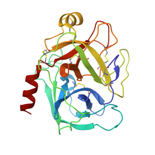
    Hydrogen bonds are key interactions determining protein-ligand binding affinity and therefore fundamental to any biological process. Unfortunately, explicit structural information about hydrogen positions and thus H-bonds in protein-ligand complexes is extremely rare and similarly the important role of water during binding remains poorly understood. Here, we report on neutron structures of trypsin determined at very high resolutions ≤1.5 Å in uncomplexed and inhibited state complemented by X-ray and thermodynamic data and computer simulations. Our structures show the precise geometry of H-bonds between protein and the inhibitors N-amidinopiperidine and benzamidine along with the dynamics of the residual solvation pattern. Prior to binding, the ligand-free binding pocket is occupied by water molecules characterized by a paucity of H-bonds and high mobility resulting in an imperfect hydration of the critical residue Asp189. This phenomenon likely constitutes a key factor fueling ligand binding via water displacement and helps improving our current view on water influencing protein-ligand recognition.


  • Organizational Affiliation

    Institut für Pharmazeutische Chemie, Philipps-Universität Marburg, Marbacher Weg 6, 35032, Marburg, Germany.


Macromolecules
Find similar proteins by:  (by identity cutoff)  |  3D Structure
Entity ID: 1
MoleculeChains Sequence LengthOrganismDetailsImage
Cationic trypsin223Bos taurusMutation(s): 0 
EC: 3.4.21.4
UniProt
Find proteins for P00760 (Bos taurus)
Explore P00760 
Go to UniProtKB:  P00760
Entity Groups  
Sequence Clusters30% Identity50% Identity70% Identity90% Identity95% Identity100% Identity
UniProt GroupP00760
Sequence Annotations
Expand
  • Reference Sequence
Small Molecules
Ligands 4 Unique
IDChains Name / Formula / InChI Key2D Diagram3D Interactions
J3I
Query on J3I

Download Ideal Coordinates CCD File 
H [auth A](2~{S})-1-[(2~{R})-2-azanyl-3-phenyl-propanoyl]-~{N}-[(1-carbamimidoylpiperidin-4-yl)methyl]pyrrolidine-2-carboxamide
C21 H32 N6 O2
MQLNFLUDIXFKTN-MSOLQXFVSA-N
SO4
Query on SO4

Download Ideal Coordinates CCD File 
C [auth A],
D [auth A],
E [auth A],
F [auth A],
G [auth A]
SULFATE ION
O4 S
QAOWNCQODCNURD-UHFFFAOYSA-L
DMS
Query on DMS

Download Ideal Coordinates CCD File 
I [auth A],
J [auth A]
DIMETHYL SULFOXIDE
C2 H6 O S
IAZDPXIOMUYVGZ-UHFFFAOYSA-N
CA
Query on CA

Download Ideal Coordinates CCD File 
B [auth A]CALCIUM ION
Ca
BHPQYMZQTOCNFJ-UHFFFAOYSA-N
Binding Affinity Annotations 
IDSourceBinding Affinity
J3I Binding MOAD:  5MNQ Kd: 3600 (nM) from 1 assay(s)
Experimental Data & Validation

Experimental Data

  • Method: X-RAY DIFFRACTION
  • Resolution: 1.34 Å
  • R-Value Free: 0.171 
  • R-Value Work: 0.137 
  • R-Value Observed: 0.139 
  • Space Group: P 21 21 21
Unit Cell:
Length ( Å )Angle ( ˚ )
a = 59.893α = 90
b = 64.076β = 90
c = 69.435γ = 90
Software Package:
Software NamePurpose
PHENIXrefinement
XDSdata reduction
XDSdata scaling
PHASERphasing
Cootmodel building

Structure Validation

View Full Validation Report


Ligand Structure Quality Assessment 


Entry History & Funding Information

Deposition Data


Funding OrganizationLocationGrant Number
European Research Council268145-DrugProfilBind

Revision History  (Full details and data files)

  • Version 1.0: 2018-01-17
    Type: Initial release
  • Version 1.1: 2018-09-12
    Changes: Data collection, Database references
  • Version 1.2: 2024-01-17
    Changes: Data collection, Database references, Refinement description