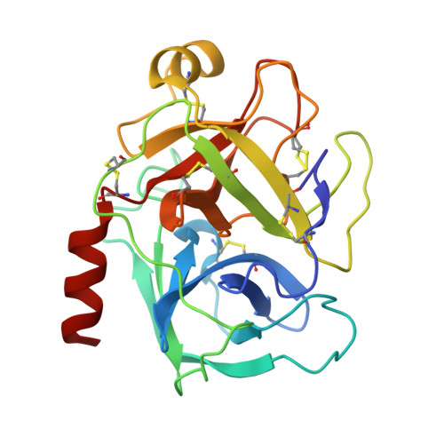
5MN1

Cationic trypsin in complex with 2-aminopyridine (deuterated sample at 100 K)


Experimental Data Snapshot

  • Method: X-RAY DIFFRACTION
  • Resolution: 0.79 Å
  • R-Value Free: 0.109 
  • R-Value Work: 0.098 
  • R-Value Observed: 0.099 

Starting Model: experimental
View more details

wwPDB Validation   3D Report Full Report


This is version 1.2 of the entry. See complete history


Literature

Charges Shift Protonation: Neutron Diffraction Reveals that Aniline and 2-Aminopyridine Become Protonated Upon Binding to Trypsin.

Schiebel, J.Gaspari, R.Sandner, A.Ngo, K.Gerber, H.D.Cavalli, A.Ostermann, A.Heine, A.Klebe, G.

(2017) Angew Chem Int Ed Engl 56: 4887-4890

  • DOI: https://doi.org/10.1002/anie.201701038
  • Primary Citation of Related Structures:  
    5MN1, 5MNA, 5MNB, 5MNC, 5MNX, 5MNY, 5MON, 5MOO

  • PubMed Abstract: 

    Hydrogen atoms play a key role in protein-ligand recognition. They determine the quality of established H-bonding networks and define the protonation of bound ligands. Structural visualization of H atoms by X-ray crystallography is rarely possible. We used neutron diffraction to determine the positions of the hydrogen atoms in the ligands aniline and 2-aminopyridine bound to the archetypical serine protease trypsin. The resulting structures show the best resolution so far achieved for proteins larger than 100 residues and allow an accurate description of the protonation states and interactions with nearby water molecules. Despite its low pK a of 4.6 and a large distance of 3.6 Å to the charged Asp189 at the bottom of the S1 pocket, the amino group of aniline becomes protonated, whereas in 2-aminopyridine, the pyridine nitrogen picks up the proton although its amino group is 1.6 Å closer to Asp189. Therefore, apart from charge-charge distances, tautomer stability is decisive for the resulting binding poses, an aspect that is pivotal for predicting correct binding.


  • Organizational Affiliation

    Institut für Pharmazeutische Chemie, Philipps-Universität Marburg, Marbacher Weg 6, 35032, Marburg, Germany.


Macromolecules
Find similar proteins by:  (by identity cutoff)  |  3D Structure
Entity ID: 1
MoleculeChains Sequence LengthOrganismDetailsImage
Cationic trypsin223Bos taurusMutation(s): 0 
EC: 3.4.21.4
UniProt
Find proteins for P00760 (Bos taurus)
Explore P00760 
Go to UniProtKB:  P00760
Entity Groups  
Sequence Clusters30% Identity50% Identity70% Identity90% Identity95% Identity100% Identity
UniProt GroupP00760
Sequence Annotations
Expand
  • Reference Sequence
Experimental Data & Validation

Experimental Data

  • Method: X-RAY DIFFRACTION
  • Resolution: 0.79 Å
  • R-Value Free: 0.109 
  • R-Value Work: 0.098 
  • R-Value Observed: 0.099 
  • Space Group: P 21 21 21
Unit Cell:
Length ( Å )Angle ( ˚ )
a = 54.692α = 90
b = 58.269β = 90
c = 66.936γ = 90
Software Package:
Software NamePurpose
PHENIXrefinement
XDSdata reduction
XDSdata scaling
PHASERphasing
Cootmodel building

Structure Validation

View Full Validation Report


Entry History & Funding Information

Deposition Data


Funding OrganizationLocationGrant Number
European Research CouncilGermany268145-DrugProfilBind

Revision History  (Full details and data files)

  • Version 1.0: 2017-05-24
    Type: Initial release
  • Version 1.1: 2018-03-14
    Changes: Author supporting evidence, Data collection, Refinement description
  • Version 1.2: 2024-01-17
    Changes: Data collection, Database references, Refinement description